The work, led by Yifan Xie, Qi Cheng, Li Wang and colleagues with Huaxiang Wu as senior author, was published online on 22 October 2026 as a peer-reviewed, accepted manuscript carrying a permanent DOI, with a final version of record to follow.
Rheumatoid arthritis is a chronic autoimmune disease in which fibroblast-like synoviocytes, the cells that line the joint, proliferate and activate abnormally. The study’s authors note that this pathological process is closely associated with ferroptosis, a cell death mechanism characterized by the accumulation of lipid peroxides, and their research was designed to clarify how artesunate might exploit that connection.
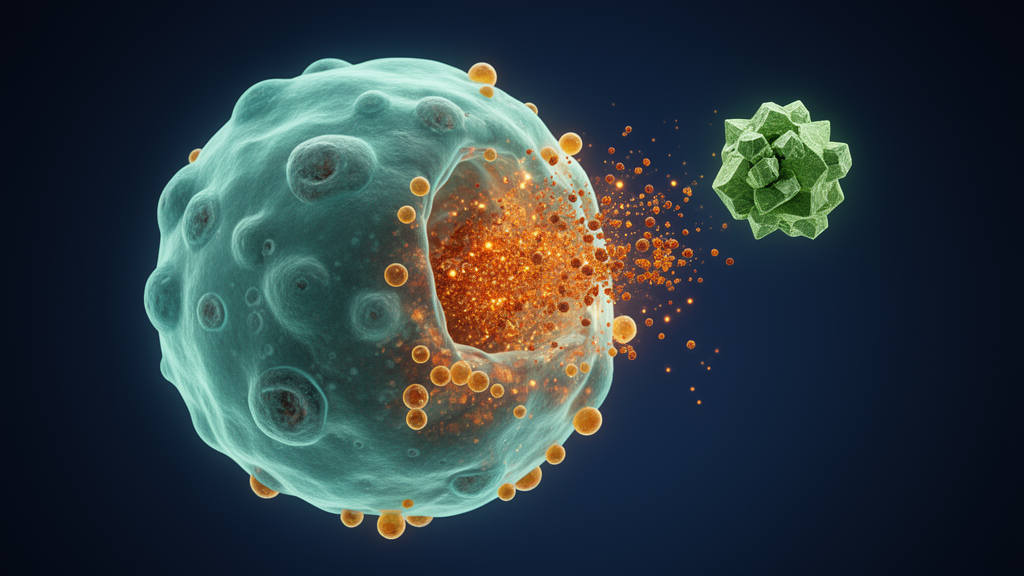
In a series of laboratory experiments, the team treated rheumatoid arthritis synovial fibroblasts, or RASFs, as well as synovial fibroblasts derived from osteoarthritis patients, with artesunate. They measured cell proliferation, invasion and migration, and found that the drug inhibited cytokine secretion, proliferation and migration in these cells. At the same time, artesunate promoted ferroptosis: the treated cells showed increased levels of reactive oxygen species, malondialdehyde and lipid hydroperoxides, alongside decreased levels of glutathione and the ferroptosis-defending enzyme GPX4.
The causal link between ferroptosis and the drug’s effects on cell activation was tested directly. When ferroptosis was blocked, the suppressive effects of artesunate on cellular activation, including cytokine release, proliferation and migration, were significantly reversed. This reversal suggests that the induction of ferroptosis is not merely a side effect of treatment but a mechanistic component of how artesunate restrains the aggressive behavior of synovial fibroblasts in this model system.
To identify the signaling pathway involved, the researchers performed RNA sequencing to analyze differentially enriched signaling pathways and used molecular docking to predict potential protein targets. According to the study, artesunate downregulated GPX4 expression through suppression of JAK1/STAT3 signaling, thereby removing a key cellular defense against lipid peroxidation and allowing ferroptosis to proceed. Key findings were validated by Western blotting and ChIP-qPCR, and transmission electron microscopy was used to examine cell morphology consistent with ferroptosis.
The team then moved from cell culture to an animal model. They established a collagen-induced arthritis mouse model, a widely used experimental approximation of rheumatoid arthritis, and compared arthritis symptoms, pathological changes and immunological responses between artesunate-treated and control groups. In these experiments, artesunate ameliorated arthritis severity, as evidenced by reduced arthritis scores and reduced joint swelling, along with improved histopathological parameters and decreased cytokine levels in joint supernatants and serum.
To probe the mechanism in vivo, the researchers isolated primary synovial fibroblasts from the synovial tissue of the arthritic mice. Part of the therapeutic effect, the authors conclude, may be attributed to the induction of ferroptosis in synovial fibroblasts within the affected joints, linking the cellular mechanism observed in vitro to the improvements seen in the living animals.
The study’s conclusion is that artesunate inhibits the activation of synovial fibroblasts by inducing ferroptosis through the JAK1/STAT3-GPX4 signaling pathway, thereby attenuating joint damage. The authors summarize the work in three highlights: in RASFs, artesunate inhibits cell activation, including cytokine secretion, proliferation and migration; it does so by inducing ferroptosis through the JAK1/STAT3 signaling pathway; and in animal models, artesunate attenuates collagen-induced arthritis by inducing the ferroptosis of synovial fibroblasts from arthritic mice.
Several caveats are worth keeping in view. The findings come from cell culture experiments and a collagen-induced arthritis mouse model, not from human clinical trials, so the results demonstrate therapeutic potential in experimental systems rather than established efficacy in patients. The authors themselves state that only part of the therapeutic effect may be attributed to ferroptosis induction in joint synovial fibroblasts, indicating that other mechanisms may contribute. In addition, the published version is an early-release, peer-reviewed accepted manuscript that remains subject to further edits before the final version of record appears.
Nevertheless, the study adds to a growing body of work connecting ferroptosis to rheumatoid arthritis biology and positions the JAK1/STAT3-GPX4 axis as a mechanistic link between an artemisinin-derived drug and the behavior of the synovial cells that drive joint damage.
Subject of Research: Preclinical research on artesunate inducing ferroptosis via the JAK1/STAT3-GPX4 pathway to attenuate rheumatoid arthritis
Article Title: Artesunate attenuates rheumatoid arthritis by inducing ferroptosis through the JAK1/STAT3 pathway
Article References: Xie, Y., Cheng, Q., Wang, L., Zhou, F., Xu, M., Zhu, L., Wang, F., Tong, S., Li, X., Lu, K., Du, Y., & Wu, H. (2026). Artesunate attenuates rheumatoid arthritis by inducing ferroptosis through the JAK1/STAT3 pathway. Arthritis Research & Therapy. https://doi.org/10.1186/s13075-026-03933-4
Image Credits: AI Generated
DOI: 10.1186/s13075-026-03933-4
Keywords: rheumatoid arthritis, artesunate, ferroptosis, JAK1/STAT3, GPX4, synovial fibroblasts, collagen-induced arthritis, artemisinin, preclinical study, oxidative stress, attenuates, rheumatoid
News Source: Ophelia Keating. (October 29, 2026). Artesunate Shows Promise Against Rheumatoid Arthritis by Triggering Ferroptosis in Synovial Cells. Scienmag.