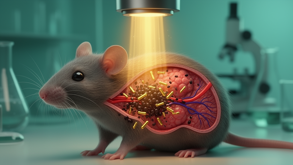
Researchers have built a single gold nanorod particle that does four jobs at once: it homes in on HER2-positive breast cancer cells, glows for near-infrared imaging, heats up under laser light to cook tumor tissue, and carries the chemotherapy drug doxorubicin. In mice bearing human SK-BR-3 breast tumors, a single injection of these particles followed by ten minutes of near-infrared irradiation made tumors vanish completely, with no measurable regrowth during a 28-day observation period. The study, published in Discover Nano, describes the platform as HER-DOX-PEG-GNRs and reports both striking efficacy and carefully acknowledged limitations.
The construction began with seed-mediated synthesis of gold nanorods, which were stripped of excess CTAB surfactant and coated with thiol-poly(ethylene glycol)-carboxylic acid. Doxorubicin was then attached covalently through EDC/NHS chemistry, forming amide bonds between PEG carboxyl groups and the drug’s primary amine. Finally, a cysteine-tagged protein G was immobilized on the gold surface through thiol-gold bonds, and trastuzumab (Herceptin) was anchored via protein G’s affinity for the antibody’s Fc region. Quantitative analysis showed the final formulation contained roughly 2.1 percent doxorubicin and 4.2 percent antibody by mass.
Each functionalization step left a measurable fingerprint. Hydrodynamic diameter grew from 54.3 nanometers for the starting CTAB-coated rods to 93.4 nanometers for the finished construct, while the zeta potential swung from strongly positive to negative after PEGylation and back to positive after drug and antibody loading. Ultraviolet-visible spectroscopy confirmed a strong longitudinal plasmon band near 805 nanometers, the optical window needed for deep tissue penetration, and transmission electron microscopy showed rods averaging 55 by 9 nanometers. In serum and cell culture medium at 37 degrees Celsius, the particles showed only modest size increases over 72 hours without substantial aggregation.
The photothermal performance proved robust. Under an 808-nanometer laser, the nanorods generated localized heat with a calculated photothermal conversion efficiency of approximately 43.3 percent, comparable to other high-performing gold nanorod systems, and repeated on-off laser cycles produced no attenuation of the heating response. In tumor-bearing mice, irradiation of the tumor region at 1.96 watts per square centimeter raised intratumoral temperature to roughly 50 degrees Celsius in animals that received the targeted particles, compared with about 41.5 degrees in mice given non-targeted rods, a difference the authors attribute to the higher tumor accumulation of the antibody-functionalized version.
That accumulation difference was substantial. Seventy-two hours after intravenous injection, inductively coupled plasma mass spectrometry recovered 6.2 percent of the injected gold dose from tumors in the targeted group versus 2.3 percent in the non-targeted group, an approximately 2.7-fold enhancement. The authors are explicit, however, that this cannot be claimed as definitive receptor-specific targeting: no HER2-negative tumor model and no receptor-blocking experiments were performed, and the particles’ positive surface charge after functionalization may itself promote nonspecific cellular association and passive enhanced permeability and retention effects. In vitro, semi-quantitative electron microscopy showed more intracellular rods in HER2-overexpressing SK-BR-3 cells treated with targeted particles than with non-targeted ones, though the difference did not reach statistical significance, and the pattern was less apparent in HER2-low MCF-7 cells.
Drug release behaved differently at different acidities. In dialysis experiments, the conjugates released more doxorubicin-associated material at pH 6.8, typical of the tumor microenvironment, than at the physiological pH 7.4. The authors caution that because doxorubicin was attached through a relatively acid-stable amide bond, the molecular basis of this pH-facilitated release remains unexplained, and the spectrophotometric assay measures doxorubicin-associated absorbance rather than confirming the chemical identity of released species. Qualitative fluorescence imaging after local injection suggested greater doxorubicin-associated signal spreading within tumor tissue than in muscle over 24 hours, though this experiment did not model systemic delivery.
The therapeutic results were the study’s centerpiece. Seven treatment groups of five mice each received saline, free doxorubicin, free trastuzumab, or nanoparticle formulations with or without irradiation. Free drugs and the complete nanoplatform without laser slowed tumor progression but did not eliminate tumors. Photothermal treatment with drug-free targeted rods shrank tumors by about 63 percent by day 14, but residual cells repopulated the tumor, which regrew to roughly 15 percent above its starting volume by day 28. Only the full combination, targeted rods carrying doxorubicin plus irradiation, produced complete regression in all animals by day 5, with zero measurable tumor volume on day 28. Prespecified statistical comparisons confirmed the day 28 advantage over the three closest comparator groups, and a longitudinal mixed-effects model showed tumor trajectories differing significantly among groups.
Histology corroborated the clinical picture. Tumors from the combination group showed PCNA-positive nuclei falling from 41.6 percent in controls to 8.5 percent, while TUNEL-positive area rose from 14.5 to 61.8 percent, consistent with suppressed proliferation and extensive DNA fragmentation and cell death. In cultured SK-BR-3 cells, the combination reduced viability by an additional 42 percent relative to the same particles without irradiation, and photothermal treatment alone accounted for roughly an 18 percent reduction. The cells also showed a delayed heat-shock response, with HSP27 significantly elevated at 24 hours after irradiation while HSP70 remained unchanged; the authors note that whether this induction contributed to thermotolerance or therapeutic outcome was not established.
Preliminary safety signals were reassuring but limited. Zebrafish embryos exposed to the particles at 6 to 24 micrograms per milliliter for 72 hours showed no significant changes in survival, hatching, heart rate, or gross morphology. Over 28 days in mice, the treated animals showed no obvious clinical signs, no marked weight loss, and no pathological abnormalities in major organs by hematoxylin and eosin staining. Most hematological and serum biochemical parameters were unchanged, although white blood cell count and blood urea nitrogen were modestly but significantly elevated, remaining within reported reference ranges. Gold accumulated predominantly in liver and spleen, consistent with reticuloendothelial sequestration, and tissue levels declined by day 28, though residual gold remained detectable and clearance kinetics were not fully characterized.
The authors frame the advance as integration rather than any single breakthrough: a solution-phase, batch-amenable fabrication route combining antibody targeting, NIR imaging, localized heating, and drug delivery in one construct, improving on their earlier substrate-deposited half-shell design. They equally emphasize what remains undone, including HER2-negative and blocking controls, dose-response and formal synergy analysis, longer follow-up to assess recurrence and metastasis, and extended safety and clearance studies. The free-drug comparator groups were not equivalent-dose comparisons, so superiority over conventional chemotherapy cannot be claimed from these regimens. What the study does establish is that, under its tested conditions, a single multifunctional gold nanorod platform achieved complete and durable tumor regression in a HER2-overexpressing xenograft model, a result that will push the field toward rigorous validation of receptor-specific targeting and long-term biocompatibility.
Subject of Research: HER2-targeted gold nanorods for imaging-guided chemo-photothermal therapy of HER2-positive breast cancer
Article Title: HER2-targeted multifunctional gold nanorods for imaging-guided combined chemotherapy and photothermal therapy
Article References: Lee, S.-M., Park, H., Han, J., Park, Y.-B., & Yoo, K.-H. (2026). HER2-targeted multifunctional gold nanorods for imaging-guided combined chemotherapy and photothermal therapy. Discover Nano, 21(1), Article 558. https://doi.org/10.1186/s11671-026-05059-7
Image Credits: AI Generated
DOI: 10.1186/s11671-026-05059-7
Keywords: gold nanorods, HER2, doxorubicin, photothermal therapy, chemotherapy, near-infrared imaging, drug delivery, breast cancer, xenograft, nanomedicine, trastuzumab, tumor regression
News Source: Nathaniel Bowman. (October 29, 2026). Antibody-Coated Gold Nanorods Erase Breast Tumors in Mice Under Near-Infrared Light. Scienmag.