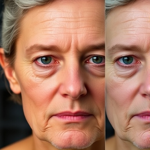
A groundbreaking study emerging from the collaborative research team at Mass General Brigham is pushing the frontiers of oncology and artificial intelligence with an innovative tool named FaceAge. This AI-driven technology, originally devised to estimate biological age from a single photograph, is now revealing unprecedented capabilities when applied to longitudinal facial image data. By analyzing changes in biological age over time, determined via serial photographs, scientists have demonstrated an enhanced capacity to predict cancer patient outcomes more accurately than with conventional methods.
The foundation of FaceAge lies in deep learning algorithms capable of deciphering complex facial features associated with biological aging. Unlike chronological age, which is simply the time elapsed since birth, biological age reflects physiological wear, cellular damage, and the cumulative burden of disease processes. By converting facial attributes into a biological age estimate, FaceAge offers a quantifiable and non-invasive biomarker that captures an individual’s health trajectory with remarkable sensitivity.
In their latest study, published in the prestigious journal Nature Communications, researchers examined a cohort of 2,279 cancer patients undergoing multiple radiation therapy sessions at Brigham and Women’s Hospital. Each participant had at least two facial photographs taken at different periods throughout their treatment timeline. By comparing biological ages derived from these sequential images, the team formulated a new metric named Face Aging Rate (FAR), which quantifies how quickly a patient’s biological age changes relative to chronological time.
The results were both striking and clinically significant. On average, patients’ facial biological aging advanced 40% faster than their actual chronological age over time, underscoring the toll that cancer and its treatment exert on physiological systems. Crucially, an accelerated Face Aging Rate was robustly associated with a diminished probability of survival. This correlation was particularly pronounced when the interval between photographs spanned two years or more, highlighting the value of capturing dynamic, longitudinal health data.
Face Aging Rate complements another metric used in the study, FaceAge Deviation (FAD), which assesses how biologically old or young a patient appears compared to their chronological age at a single time point. Patients who exhibited both a high FaceAge Deviation and an elevated Face Aging Rate experienced the poorest survival outcomes, revealing the synergy between static and dynamic biomarkers in characterizing health status. However, the longitudinal nature of FAR emerged as a more stable and reliable predictor over extended periods than single timepoint assessments.
The implications of these findings are far-reaching. According to Dr. Raymond Mak, co-senior author and radiation oncologist at Mass General Brigham Cancer Institute, the ability to derive a Face Aging Rate from routine clinical photographs offers a near real-time window into a patient’s evolving health. Such continuous monitoring may refine personalized treatment strategies, optimize follow-up schedules, and empower physicians to more accurately counsel patients regarding prognosis.
The technological sophistication behind FaceAge relies on advanced computational modeling and machine learning frameworks. These systems have been trained on large datasets encompassing diverse facial images and demographic profiles, enabling the AI to disentangle subtle phenotypic markers linked to cellular senescence, inflammation, and treatment-induced physiological stress. This approach transcends traditional biomarkers that often require invasive tissue sampling or costly biochemical assays, providing a scalable, accessible alternative.
Building on prior research, which demonstrated that patients with cancer typically appear approximately five years older biologically than their chronological age, the current study delves deeper into temporal changes in aging patterns. The data analytics involved meticulous processing of facial feature vectors extracted from photographs and calculation of the rate of biological aging per unit time. Such granular analysis allowed for quantifiable insights into how cancer progression and therapeutic interventions impact systemic aging mechanisms.
The broad applicability of FaceAge extends beyond oncology. Co-author Dr. Hugo Aerts, director of the Artificial Intelligence in Medicine program at Mass General Brigham, envisions potential prognostic use in various chronic diseases and even in monitoring general population health. The scalable, non-invasive nature of the tool makes it an attractive candidate for widespread clinical adoption, especially as digital health infrastructure integrates photometric data capture.
Notably, the research team has made strides toward public engagement by launching an IRB-approved web portal where individuals can upload selfies to receive FaceAge assessments. This platform not only democratizes access to health insights but also catalyzes further optimization and validation of the AI algorithm with diverse and expansive datasets, fostering translational progress.
The study’s robustness is underscored by additional research published in the Journal of the National Cancer Institute, where FaceAge was applied to over 24,500 older cancer patients receiving radiation therapy. The findings aligned with prior observations: older FaceAge estimates correlated with worse survival, reinforcing the biomarker’s prognostic validity across large populations.
From a clinical perspective, the integration of Face Aging Rate assessment into routine oncology practice would represent a paradigm shift. Real-time tracking of biological aging could enable oncologists to tailor therapeutic intensity dynamically, balancing efficacy with tolerability to improve overall outcomes. Moreover, this strategy holds promise for personalizing survivorship care by identifying patients at risk of heightened physiological decline warranting closer follow-up.
Despite its promise, the researchers acknowledge that further investigation is essential to validate FaceAge and FAR across more diverse demographic and clinical contexts. Future prospective clinical trials will be critical to ascertain utility and establish standardized protocols for implementation. Ongoing interdisciplinary collaboration will continue to refine the AI algorithms, incorporating novel biomarkers and multimodal data streams to augment predictive accuracy.
In sum, the Mass General Brigham research team’s pioneering work vividly illustrates the transformative potential of AI-driven biometrics in medicine. By harnessing facial aging dynamics, FaceAge and Face Aging Rate emerge as compelling, cost-effective biomarkers that could revolutionize cancer prognosis and personal health monitoring. This fusion of technology, clinical insight, and patient engagement paves the way toward a future where non-invasive, personalized health analytics guide therapeutic decisions with unprecedented precision.
Subject of Research: People
Article Title: Face aging rate quantifies change in biological age to predict cancer outcomes
News Publication Date: 28-Apr-2026
Web References:
https://faceage.bwh.harvard.edu
https://www.massgeneralbrigham.org/en/about/newsroom/press-releases/ai-face-photos-tool-estimate-age-predict-cancer-outcomes
https://www.nature.com/articles/s41467-025-66758-w
https://academic.oup.com/jnci/advance-article-abstract/doi/10.1093/jnci/djaf323/8328045?redirectedFrom=fulltext&login=false
References:
Haugg, F. et al. “Face aging rate quantifies change in biological age to predict cancer outcomes” Nature Communications DOI: 10.1038/s41467-025-66758-w
Keywords: Artificial intelligence, Machine learning, Cancer, Oncology
Tags: AI in personalized cancer treatmentAI-driven biological age estimationbiological age vs chronological agecancer patient health trajectory monitoringdeep learning for facial biomarker analysisFaceAge technology in oncologyfacial features indicating physiological aginglongitudinal facial image data in cancernon-invasive cancer prognosis toolspredictive modeling for cancer outcomesprognostic cancer biomarkers from photosradiation therapy patient biomarker study

