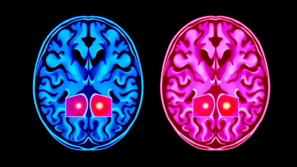
Functional magnetic resonance imaging has long promised a window into the living brain, but turning thousands of noisy, four-dimensional measurements into a clean clinical verdict has remained one of the hardest problems in medical artificial intelligence. A new study published in the journal Complex & Intelligent Systems reports a deep learning framework that tackles the problem by looking at brain function from two complementary directions at the same time, and it achieves striking results: 99.36 percent accuracy and an area under the receiver operating characteristic curve of 0.991 in classifying neurological disease severity from fMRI data.
The work, led by C. S. Smitha and S Sathiya of Annamalai University in Tamilnadu, India, together with T Soumya of the College of Engineering Muttathara in Thiruvananthapuram, Kerala, focuses on three of the most burdensome neurological conditions: Alzheimer’s disease, epilepsy, and Parkinson’s disease. Each of these disorders is marked by subtle, progressive changes in how brain regions activate and communicate with one another. Catching those changes early, and grading them accurately, matters enormously for clinical decision-making and personalized treatment planning, yet the task has stubbornly resisted automation because fMRI data are extremely high-dimensional, brain connectivity patterns shift from moment to moment, and no two brains are wired quite the same way.
The researchers’ answer is a dual-stream architecture, meaning the model processes the same input through two parallel computational pathways before merging what each has learned. The first stream, called the AdaBelief Boosted Contrastive DenseConvo net, or ABCD Net, is built on a 3D DenseNet backbone, a convolutional architecture in which each layer receives feature maps from all preceding layers, encouraging rich reuse of spatial information across the volumetric brain image. On top of that backbone, the team added Temporal Contrastive Learning, a technique that teaches the network to distinguish meaningful changes in neural activity over time from irrelevant fluctuations, and ConvLSTM fusion, which blends convolutional feature extraction with long short-term memory cells that can retain information across successive time frames.
Where the first stream asks what is happening inside the brain’s tissue, the second asks how the brain’s regions are talking to each other as the scan unfolds. This stream, named the Dynamic Slepian Gated Recurrent Transformer, or DynSGRT, constructs windowed representations of dynamic functional connectivity, the time-varying statistical relationships between activity in different brain areas. Rather than treating connectivity as a single static snapshot, the model slices the scanning session into windows and builds a graph for each one, with brain regions as nodes and their temporal correlations as edges. Slepian basis functions, which are optimally concentrated in both time and frequency, help represent these dynamic signals compactly, while gated recurrent units and attention mechanisms track how the connectivity patterns evolve.
Those windowed connectivity graphs are then handed to a third component, the Hierarchically differentiated temporal-evolving graph neural network, or HD-TEG Net. Graph neural networks are a class of deep learning models designed to operate on data structured as networks rather than grids, and they are a natural fit for brain connectivity, which is inherently a network problem. HD-TEG Net applies graph pooling, a method for progressively coarsening a graph into higher-level summaries, and temporal evolution modeling, which captures how the pooled graph representations change from one window to the next. In effect, the second stream learns a compressed story of how the brain’s communication network reorganizes itself over the course of a scan.
Bringing the two stories together is where the framework earns its multimodal label. The researchers use a Cross-modal dual-head attentive concatenation strategy, in which two attention heads weigh the contributions of the spatial-temporal stream and the connectivity stream before their features are concatenated into a single joint representation. This lets the classifier learn, for each case, whether structural activity patterns or connectivity dynamics carry more diagnostic signal, or how best to combine them. The fused representation is then classified by a Chicken swarm optimized DEformer, a dynamic embedding transformer whose training is guided by a bio-inspired optimization algorithm modeled on the hierarchical foraging search of chicken flocks. The optimization step is intended to improve generalization and help the model avoid poor local minima during training, a common pitfall for deep networks on limited medical datasets.
The evaluation design deserves particular attention, because inflated results are a chronic problem in machine learning applied to brain imaging. The team assembled a balanced benchmark of 800 subjects drawn from the NeuroVault and NITRC public repositories, with 200 subjects each in four groups: Alzheimer’s disease, Parkinson’s disease, epilepsy, and healthy normal controls. Data were split in a subject-independent 70:30 train-test ratio, meaning that no individual contributing training data ever appeared in the test set, and the pipeline was further validated with five-fold cross-validation. All headline results were computed on completely unseen testing subjects, a protocol designed to ensure that the reported performance reflects genuine diagnostic capability rather than memorization of individual scan signatures.
On that benchmark, the framework reached 99.36 percent accuracy with an AUC-ROC of 0.991, figures the authors report as significantly outperforming both baseline models and previously published approaches in the literature. An AUC-ROC near 0.99 means that, for almost any threshold a clinician might choose, the model separates diseased from healthy or distinguishes severity levels with near-perfect trade-offs between sensitivity and specificity. For disorders like Alzheimer’s and Parkinson’s, where severity grading can guide when to escalate therapy, and for epilepsy, where characterizing abnormal activity patterns informs surgical and pharmacological decisions, a reliable automated second opinion could meaningfully change clinical workflows.
Why might the dual-stream design matter so much? Neuroscientists have increasingly recognized that disease signatures live in both places: in the raw spatiotemporal patterns of neural activity and in the way connectivity between regions degrades or reorganizes as pathology advances. Alzheimer’s disease, for example, is associated with progressive disruption of large-scale network communication, while epilepsy involves abnormal synchrony that can emerge and dissipate on rapid timescales. A single-stream model sees only one side of that picture. By fusing a volumetric convolutional view of the brain with a graph-based view of its evolving functional architecture, the framework bridges what the authors describe as spatiotemporal and connectivity-based perspectives of brain function, extracting complementary evidence that neither view alone provides.
The study, published open access on 6 October 2026, arrives amid a broader surge of interest in applying graph neural networks, transformers, and hybrid bio-inspired optimizers to neuroimaging, and it suggests that the next generation of diagnostic tools may increasingly be multimodal even when the input is a single imaging modality. The authors declare no competing interests and report no external funding for the work. Significant hurdles remain before such systems reach the clinic, including validation on larger and more diverse populations, robustness to differences in scanner hardware and acquisition protocols, and prospective testing in real diagnostic settings. But the message of the study is clear: when a model is built to watch both what the brain does and how its parts converse, the faint fingerprints of neurological disease become far easier to read, and the long-sought bridge between fMRI research and bedside decision support moves a measurable step closer.
Subject of Research: Deep learning classification of neurological disease severity from fMRI using spatiotemporal and dynamic functional connectivity features
Article Title: Dual stream spatiotemporal feature learning framework for neurological disease severity level classification using fMRI data
Article References: Smitha, C. S., Sathiya, S., & Soumya, T. (2026). Dual stream spatiotemporal feature learning framework for neurological disease severity level classification using fMRI data. Complex & Intelligent Systems. https://doi.org/10.1007/s40747-026-02527-6
Image Credits: AI Generated
DOI: 10.1007/s40747-026-02527-6
Keywords: fMRI, neurological disorders, Alzheimer's disease, Parkinson's disease, epilepsy, deep learning, dynamic functional connectivity, graph neural network, 3D DenseNet, transformer, disease severity classification, brain connectivity
News Source: Cassandra Pierce. (October 6, 2026). AI Reads Brain Scans Two Ways at Once to Gauge Neurological Disease Severity. Scienmag.