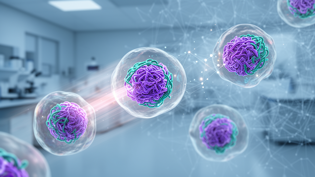
Deep inside the nucleus of every blood stem cell lies a record of how old that cell really is. It is not written in letters of DNA sequence, but in the three-dimensional architecture of chromatin, the tangled complex of DNA and proteins that packages the genome and decides which genes are accessible and which are silenced. A research team from the Bellvitge Biomedical Research Institute (IDIBELL), the Barcelona Supercomputing Center – Centro Nacional de Supercomputación (BSC-CNS) and the Barcelona Institute for Global Health (ISGlobal) has now shown that this architectural record can be read automatically by artificial intelligence, using nothing more than a standard fluorescence image of a stained nucleus. Their tool, called ChromAgeNet, is described in the journal Aging Cell and could change how researchers screen for interventions that preserve or restore the function of aging blood-forming cells.
The study was co-led by Dr Maria Carolina Florian, a researcher in the Regenerative Medicine program at IDIBELL and ICREA Research Professor, and Dr Paula Petrone, a researcher at BSC-CNS and ISGlobal, a centre supported by the la Caixa Foundation. The work formed a central part of the doctoral thesis of Pablo Iañez, a researcher at ISGlobal, and combined expertise in stem cell biology, aging, image analysis and machine learning. Their starting point was a well-known clinical and biological problem: as the body ages, the hematopoietic system, the network of organs and tissues responsible for producing blood cells, progressively loses its capacity to maintain adequate blood cell production. Measuring this decline at the level of individual stem cells is essential for understanding hematopoietic aging and for testing strategies designed to slow or reverse it.
Chromatin is the material that makes this measurement possible. It consists mainly of DNA wrapped around proteins, and its spatial organization within the nucleus regulates gene activity, ultimately determining cell identity and function. As stem cells age, this organization changes, but the changes are subtle, heterogeneous and often invisible to a trained observer looking through a microscope. That is precisely where the new model excels. ChromAgeNet is a convolutional neural network, a class of artificial intelligence model designed to analyze images, and it was trained to distinguish young from aged hematopoietic stem cells based solely on the appearance of the nucleus.
To build and train the model, the researchers used three-dimensional microscopy images of mouse hematopoietic stem cell nuclei stained with DAPI, a simple, inexpensive and widely used fluorescent dye that binds to DNA and reveals the distribution of chromatin density inside the nucleus. The choice of DAPI is significant for practical reasons. Because it is a low-cost stain that can be incorporated easily into existing microscopy protocols, the approach does not require exotic reagents or specialized sample preparation, which makes it realistic to deploy in high-throughput microscopy workflows where thousands or millions of samples must be processed automatically.
The performance results are striking. Based on the appearance of the nucleus alone, ChromAgeNet demonstrated a 77 percent probability of correctly sorting cells into two groups, young versus aged. Remarkably, this exceeded the performance of a machine learning model built on chromatin features that the researchers themselves had previously defined by hand. In other words, the deep learning system extracted predictive information from the images that went beyond the features human experts had considered informative. This is a recurring theme in modern image-based biology: neural networks can detect combinations of visual cues that no single engineered feature captures, and here those cues turned out to encode biological age.
One of the most important aspects of the work is that the aging-associated differences are not necessarily visible to the naked eye in the microscopic images. Stem cell aging is a heterogeneous process, meaning that different cells age in different ways and to different degrees, and changes in nuclear architecture can be extremely subtle. ChromAgeNet nevertheless manages to identify combinations of spatial chromatin features that carry information about the aging state of individual cells. This transforms a subjective, qualitative impression of an aged nucleus into a quantitative, scalable measurement that can be applied consistently across large cell populations.
Crucially, the team did not stop at classification. They analyzed the model’s behaviour to determine which specific features of the images were most helpful in distinguishing young cells from aged cells. This interpretability analysis identified several predictive features of the age-associated state, including chromatin entropy, a measure of the disorder or variability in chromatin organization; heterochromatin, the densely packed and generally gene-poor form of chromatin, located at the periphery of the nucleus; and certain chromatin condensates, compact clusters of the DNA-protein complex. Knowing which elements of DNA organization the model relies on allows researchers to go beyond a simple young-versus-old label and gain mechanistic insight into the nuclear architecture of aging.
This mechanistic dimension positions ChromAgeNet as a complement to existing biological age biomarkers rather than a replacement for them. The best known of these are the so-called epigenetic clocks, which estimate biological age from chemical modifications to DNA, particularly methylation patterns. Epigenetic clocks require sequencing-based measurements, whereas ChromAgeNet requires only a stained image, so the two approaches interrogate different layers of the aging process. Combining image-derived chromatin architecture with molecular biomarkers could yield a richer, multi-dimensional picture of how a given stem cell is aging and how it responds to experimental interventions.
The most forward-looking application explored in the study is screening for rejuvenation strategies. As a proof of concept, the researchers applied ChromAgeNet to aged hematopoietic stem cells that had been treated with different epigenetic drugs, asking whether the treatments produced changes in chromatin organization compatible with a younger nuclear state. The authors are careful about what this does and does not show. The results do not demonstrate that the treatments functionally rejuvenated the cells, but they do establish the potential of ChromAgeNet as a tool to detect age-associated chromatin changes in response to interventions. That distinction matters: a model that can flag candidate compounds for closer functional testing accelerates discovery without overclaiming therapeutic effect.
The combination of cheap staining, a small number of model parameters and compatibility with high-throughput microscopy makes the platform unusually scalable. In principle, the same pipeline could be used to study large libraries of compounds, rapidly identifying those that shift the nuclear architecture of aged stem cells toward a younger configuration, which would then be prioritized for deeper functional assays. To support exactly this kind of community-driven development, the researchers have released a dataset of three-dimensional images of hematopoietic stem cells together with ChromAgeNet itself. This is a meaningful contribution in its own right, because publicly available imaging datasets for hematopoietic stem cell aging are currently scarce, and shared benchmarks are what allow computational tools to be validated and improved across laboratories.
Taken together, the study demonstrates that the three-dimensional organization of DNA contains quantifiable information about the aging of blood stem cells, and that artificial intelligence can extract that information from routine microscopic images. ChromAgeNet provides a new, faster and more scalable way to study cellular aging and to explore strategies for preserving or recovering hematopoietic stem cell function. If the approach generalizes beyond mouse cells and finds adoption in screening pipelines, the humble DAPI-stained nucleus, an image type generated in cytometry and microscopy labs every day, may become one of the most informative and inexpensive windows into the biological age of the cells that keep our blood, and ultimately our bodies, running.
Subject of Research: Deep learning detection of hematopoietic stem cell aging from 3D chromatin architecture in nuclear images
Article Title: AI detects signs of aging in blood stem cells from nuclear images
Article References: AI detects signs of aging in blood stem cells from nuclear images. (n.d.). Original publication
Image Credits: AI Generated
DOI: Not provided
Keywords: artificial intelligence, chromatin, hematopoietic stem cells, aging, DAPI staining, convolutional neural network, nuclear architecture, epigenetic drugs, rejuvenation, Aging Cell, biomarkers, high-throughput microscopy
News Source: Beatrice Stafford. (October 17, 2026). AI reads aging in blood stem cells from images of nuclear DNA packaging. Scienmag.