


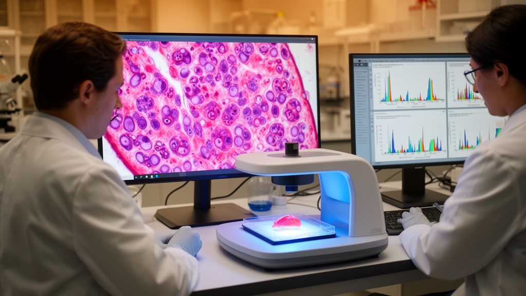
Researchers have developed a multimodal artificial intelligence framework, named XceptionAttnV1, designed to jointly process clinical metadata, medical imaging, spectral measurements and genomic features for oral cancer research. The framework addresses a well-known weakness of single-modality AI systems built on clinical photographs, histopathology or radiology alone, which offer limited predictive performance and do not support downstream treatment decisions. Its stated clinical scope is oral cavity squamous cell carcinoma and oral potentially malignant disorders.
Architecturally, the system routes each data type through a dedicated feature extractor. The imaging branch couples an Xception backbone with the Convolutional Block Attention Module, known as CBAM, to focus the network on diagnostically relevant image regions. The spectral branch, handling Raman and near-infrared measurements, is a one-dimensional convolutional network with an LSTM tail. The genomic branch uses a variational autoencoder. A cross-modal attention fusion layer then combines these representations before feeding four prediction heads: a lesion classifier, a DeepSurv-style overall-survival head, a multi-task head for recurrence and lymph-node metastasis, and a hybrid treatment-recommendation head that blends deep learning with rule-based logic.
Uncertainty handling is deliberately narrow. Only the final projection layer of the fusion module is Bayesian, implemented as a mean-field variational last layer, allowing the model to estimate epistemic uncertainty without making the entire network probabilistic. Ablation experiments reported that cross-modal attention and the Bayesian last layer each contribute separately to accuracy and to calibrated uncertainty estimates.
The reported performance figures are endpoint-specific rather than a single four-modality benchmark. On task-specific held-out partitions, the framework attained an area under the curve of 0.961 for lesion classification, an imaging-plus-clinical endpoint evaluated on oral-cavity histology cohorts. On a matched TCGA-HNSC subset for which clinical, imaging and genomic data were co-registered, it achieved a concordance index of 0.781 for overall survival, an AUC of 0.892 for lymph-node metastasis prediction, and 87.4 percent top-1 concordance with multidisciplinary tumour-board decisions. These results exceeded strong single-modality and conventional multimodal baselines evaluated under the same modality availability.
A crucial caveat shapes how these numbers should be read. Because no public archive supplies all four modalities for one oral-cancer cohort, each endpoint was evaluated only on the sources carrying the relevant modality and label, with the modality composition of every evaluated partition declared explicitly. The study states plainly that no endpoint evaluates all four modalities together, and that the framework should not be read as a validated four-modality system. It is instead a shared architecture evaluated separately on the modality-specific partitions that the available data actually support.
Where pooled sources also contained oropharyngeal or laryngeal head and neck squamous cell carcinoma records, those were analysed as a labelled sensitivity subset rather than merged silently into the oral-cancer analysis.
The authors are equally explicit about the study’s stage of development. The framework was built through a six-phase methodology, and Phase 6, prospective clinical validation, is defined as future work and has not been carried out. All results reported are retrospective, meaning the model was assessed on existing datasets rather than tested on new patients in a clinical setting. Consequently, the framework is presented as a retrospective research prototype and a structured second opinion, not as a system validated or ready for clinical deployment.
For clinicians and researchers, the work illustrates both the promise and the constraints of multimodal AI in oncology. Combining histology, CT, MRI, spectroscopy and genomics within one architecture produced strong retrospective metrics across diagnosis, survival and treatment-concordance endpoints, outperforming baselines under matched data conditions. Yet the absence of any endpoint using all four modalities simultaneously, and the lack of prospective validation, mean the reported figures describe what the architecture can achieve on available retrospective partitions, not what it would deliver at the bedside.
The publication appears in Discover Oncology as open access research, published on 28 October 2026, and is currently shared as a peer-reviewed accepted version subject to further edits before the final Version of Record.
Subject of Research: A multimodal deep learning framework for retrospective oral cancer diagnosis, survival prediction and treatment-decision concordance research.
Article Title: A multimodal attention-augmented deep learning framework for oral cancer diagnosis, prognosis and personalised treatment recommendation
Article References: Hajare, D., Deshpande, N., & Shankargouda, S. (2026). A multimodal attention-augmented deep learning framework for oral cancer diagnosis, prognosis and personalised treatment recommendation. Discover Oncology. https://doi.org/10.1007/s12672-026-06168-6
Image Credits: AI Generated
DOI: 10.1007/s12672-026-06168-6
Keywords: oral cancer, deep learning, multimodal AI, OSCC, survival prediction, lymph node metastasis, treatment recommendation, attention mechanism, Bayesian uncertainty, retrospective study, multimodal, attention-augmented
News Source: Nathaniel Bowman. (October 29, 2026). AI Framework Combines Imaging, Genomics and Spectral Data for Oral Cancer Research. Scienmag.



