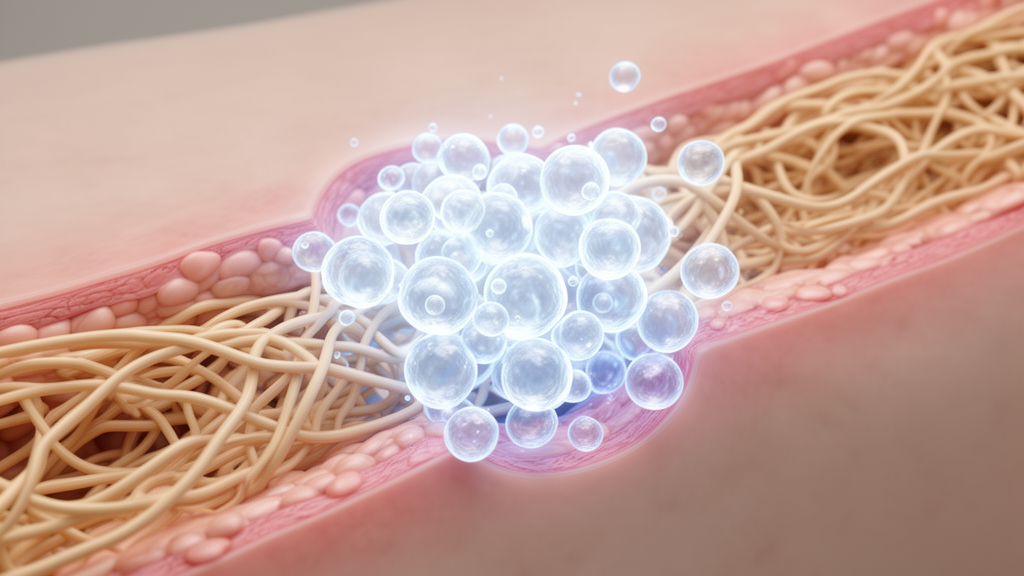
A biomaterial composite combining chitooligosaccharide with adipose-derived stem cell exosomes improved diabetic wound healing in a rat model by altering macrophage inflammatory signaling and promoting blood vessel formation, according to laboratory research published in Frontiers in Bioengineering and Biotechnology. Chronic diabetic wounds present severe clinical challenges because persistent hyperglycemia disrupts normal healing phases, locking tissue into prolonged inflammation and impairing microvascular regeneration. Researchers from Dalian University and the Affiliated Zhongshan Hospital of Dalian University developed the composite platform, designated Exo-COS, to investigate whether combining regenerative extracellular vesicles with a biocompatible chitosan derivative could bridge immune regulation and vascular repair through macrophage-mediated paracrine mechanisms.
To construct the delivery system, investigators isolated adipose-derived stem cells from six-week-old male Sprague-Dawley rats and confirmed their phenotype by demonstrating positive expression of surface markers CD49d and CD90 alongside the absence of CD34 and CD45. Exosomes isolated from third-passage stem cell culture supernatants exhibited typical nanoscale vesicle morphology under transmission electron microscopy, expressed exosomal marker proteins CD63 and TSG101, and displayed a particle diameter peak between 120 and 140 nanometers via nanoparticle tracking analysis. Mixing these vesicles with a five percent chitooligosaccharide solution at a ratio of 2.7 times 10 to the tenth particles per five milligrams of chitooligosaccharide shifted the modal hydrodynamic diameter peak upward to 150 to 160 nanometers. Confocal microscopy verified spatial colocalization of the fluorescently labeled components and demonstrated cellular internalization by vascular endothelial cells.
The therapeutic mechanism was first explored in vitro using RAW264.7 macrophages cultured under high-glucose conditions containing approximately 25 millimolar glucose to mimic the diabetic environment. Cultured macrophages were treated with chitooligosaccharide alone at 5 milligrams per milliliter, stem cell exosomes alone at 2.5 times 10 to the tenth particles per milliliter, or the Exo-COS composite at equivalent constituent concentrations. Flow cytometric analysis revealed that surface expression of the proinflammatory marker CCR7 reached its lowest level in the Exo-COS group, while intracellular levels of the reparative marker Arg-1 were highest following composite treatment. Reverse transcription quantitative PCR confirmed this immunomodulatory shift, showing marked reductions in interleukin-6 and inducible nitric oxide synthase mRNA alongside significant elevations in Arg-1 and interleukin-10 expression.
In addition to remodeling macrophage inflammatory profiles, the composite treatment significantly enhanced macrophage migratory behavior in high-glucose cultures. Scratch closure assays at 48 hours demonstrated that macrophages treated with Exo-COS achieved the highest closure rates among all tested groups, followed by exosome monotherapy and chitooligosaccharide alone. Complementary Transwell migration assays confirmed these findings, recording the greatest count of migrated macrophages traversing the membrane in the composite group.
To establish whether macrophage reprogramming functionally stimulated angiogenesis, the researchers developed an indirect conditioned-medium model. High-glucose-adapted macrophages received their designated treatments for 24 hours, followed by complete medium removal and thorough PBS washing to eliminate direct residual vesicles or biomaterial carryover. After an additional 24 hours of culture in fresh medium, macrophage-conditioned medium was harvested and applied to vascular endothelial cells pre-exposed to high glucose. Endothelial cells cultured with conditioned medium from Exo-COS-treated macrophages demonstrated superior scratch wound closure after 24 hours and the highest migration counts in Transwell chambers compared to conditioned media from untreated, chitooligosaccharide-treated, or exosome-treated cells.
Angiogenic tube formation assays on polymerized Matrigel further substantiated this indirect paracrine coupling. Endothelial cells maintained in high-glucose environments showed severely blunted capillary assembly, whereas those supplied with conditioned medium from Exo-COS-primed macrophages assembled the most extensive network architecture, generating the highest total tube lengths and the greatest number of branching nodes.
The therapeutic efficacy of the platform was evaluated in vivo using a full-thickness cutaneous wound model established in 48 diabetic Sprague-Dawley rats. Digital photographic monitoring on days 0, 7, 11, and 14 revealed that wounds treated with Exo-COS exhibited the most rapid reduction in open area, achieving the shortest overall time to complete epithelial closure compared to all other experimental groups.
Histological examinations supported the gross macroscopic healing improvements observed across the experimental timeline. Hematoxylin and eosin staining of harvested wound beds showed superior tissue reconstruction, reduced epithelial defect lengths, and more organized dermal architecture in the Exo-COS cohort. Masson’s trichrome staining demonstrated substantial collagen deposition within the regenerating dermis of composite-treated animals, whereas untreated diabetic control wounds displayed sparse, disorganized extracellular matrix fibrils. These structural evaluations confirmed that local application of the composite accelerated constructive remodeling alongside re-epithelialization.
Immunohistochemical and immunofluorescence analyses of wound tissue biopsies corroborated the in vitro mechanistic findings within living tissue. Multi-marker immunofluorescence staining demonstrated that diabetic wound beds treated with Exo-COS had significantly diminished CCR7 expression alongside increased Arg-1 staining, confirming an in vivo transition toward a reparative tissue profile. Concurrently, immunohistochemical labeling for endothelial marker CD31 revealed that Exo-COS-treated wounds established the highest density of microvessels across six examined microscopic fields. This correlation between reduced local inflammation and heightened capillary density supported the role of immune-vascular coupling in promoting tissue survival.
The authors highlighted several critical experimental limitations that contextualize their results and outline future translational priorities. While CCR7, Arg-1, interleukin-6, and interleukin-10 provided clear markers of inflammatory status, they do not encompass the full phenotypic heterogeneity of tissue macrophages in vivo. Furthermore, although conditioned-medium experiments demonstrated functional immune-endothelial communication, the study did not isolate specific paracrine cytokines, employ macrophage depletion models, or evaluate exosome release kinetics and long-term biodistribution. Additionally, while CD31-positive microvessel counts established enhanced structural neovascularization, they did not measure functional vessel perfusion or vascular maturation in the healed tissues.
Methodological protocols provided detailed specifications for primary cell processing and composite formulation. To obtain adipose-derived stem cells, harvested rodent adipose tissue was minced and digested with 0.2 percent type I collagenase at 37 degrees Celsius for one hour before passing through a 100-micrometer cell strainer. After establishing adherent cultures, exosome isolation was initiated once cells reached approximately 80 percent confluence. Conditioned medium collected after 48 hours of serum-depleted culture was cleared of debris at 3,000 g prior to exosome concentration. Nanoparticle tracking analysis confirmed size distributions, while Western blot evaluations demonstrated positive signals for CD63, CD90, and TSG101 alongside an absence of beta-actin, confirming vesicular purity. To formulate the composite, chitooligosaccharide was dissolved in culture medium at five percent, adjusted to pH 6.5 using 1.5 molar Tris-HCl, and incubated under gentle rotation with exosomal suspensions for one hour at room temperature.
The indirect macrophage-endothelial co-culture model adhered to strict preparation protocols to ensure observed endothelial responses stemmed from paracrine factors rather than direct reagent carryover. Following initial 24-hour treatments under high-glucose conditions, RAW264.7 macrophages were washed three times with phosphate-buffered saline before fresh medium was introduced for an additional 24 hours. Supernatants were subsequently harvested and subjected to sequential centrifugation at 300 g for five minutes and 2,000 g for ten minutes at 4 degrees Celsius to eliminate floating cells and debris. In downstream angiogenic assays, vascular endothelial cells pre-exposed to high glucose were treated with conditioned media diluted at a one-to-one volume ratio with fresh growth medium. Transwell assays evaluated migration by seeding 100,000 endothelial cells across eight-micrometer porous membranes, while Matrigel capillary network formation was assessed after seeding 65,000 cells per well for 24 hours.
Surgical procedures and quantitative histology followed standardized experimental parameters. The 56 male Sprague-Dawley rats, weighing between 260 and 300 grams, were obtained from Liaoning Changsheng Biological, with eight animals used for cell isolation and 48 assigned to diabetic wound studies. After establishing diabetic status and generating full-thickness skin defects, topical treatments were applied directly to the wound surface and left undisturbed for 30 minutes before bandaging. Harvested wound tissues were fixed in four percent paraformaldehyde for 48 hours before paraffin sectioning. Microscopic assessments evaluated tissue reconstruction across six randomly selected fields at 400-times magnification, measuring Masson’s trichrome collagen staining and CD31-positive microvessel counts. Statistical comparisons across groups relied on one-way or two-way analysis of variance in GraphPad Prism, supplemented by Tukey’s multiple-comparisons tests.
In discussing their findings, the authors emphasized important biological and structural nuances. Rather than describing cellular responses as complete conversions between rigid, binary M1 and M2 phenotypes, the researchers characterized the observed shifts as continuous alterations in inflammatory-reparative states, reflecting the known plasticity and heterogeneity of wound macrophages. Structurally, while confocal colocalization and hydrodynamic size shifts confirmed integration within the composite, the investigators noted that these measurements do not demonstrate covalent binding or specific molecular linkages between chitooligosaccharide and vesicular membranes. Furthermore, although chitooligosaccharide monotherapy demonstrated relatively modest immunomodulatory activity in cultured macrophages, it produced noticeable wound repair in vivo, indicating that the biomaterial independently influences surrounding fibroblasts, extracellular matrix components, or alternative wound microenvironment processes.
Subject of Research: Diabetic wound repair via biomaterial-exosome composite system
Article Title: Chitooligosaccharide–adipose-derived stem cell exosome complex promotes diabetic wound healing through macrophage-mediated immune–vascular coupling
Article References: Dong, Y., Yan, Z., Liu, B., Wang, C., Zhang, Z., & Liu, J. (2026). Chitooligosaccharide–adipose-derived stem cell exosome complex promotes diabetic wound healing through macrophage-mediated immune–vascular coupling. Frontiers in Bioengineering and Biotechnology, 14, Article 1999030. https://doi.org/10.3389/fbioe.2026.1999030
Image Credits: AI Generated
DOI: 10.3389/fbioe.2026.1999030
Keywords: diabetic wound healing, adipose-derived stem cells, exosomes, chitooligosaccharide, macrophage polarization, angiogenesis, immune-vascular coupling, extracellular vesicles, tissue reconstruction, endothelial cell migration, tube formation, Sprague-Dawley rats, CD31, arginase-1, CCR7, regenerative biomaterials
News Source: Drew Townsend. (November 19, 2026). Stem Cell Exosome Composite Accelerates Diabetic Wound Healing in Rat Model. Scienmag.