A paradigm shift is emerging in cancer research that challenges the classical genocentric view of lung carcinogenesis, which has long emphasized the random acquisition of genetic mutations as the primary driver of tumor development. Instead, a recent review synthesizes extensive evidence to propose a mechanochemical coupling framework, suggesting that the physical microenvironment of the lung—specifically pathological mechanical forces—is a major contributor to malignant progression. This framework posits that high-magnitude cyclic stretch and excessive extracellular matrix stiffening function as physical stimuli that are translated into sustained oxidative stress and genomic instability within the lung tumor microenvironment. By integrating biophysical principles with molecular oncology, this review highlights a series of mechanochemical checkpoints that may define how mechanical cues drive the lung toward a malignant phenotype, potentially offering new therapeutic targets for intercepting cancer at its mechanical roots.
The mechanical landscape of the lung is fundamentally altered during malignant transformation, transitioning from a compliant state to one characterized by pathological rigidity. Normal lung parenchyma typically exhibits a Young’s modulus between 1 and 5 kPa, providing the necessary elasticity for gas exchange and recoil. However, cancer-associated fibroblasts often mediate a desmoplastic reaction, leading to an overproduction and cross-linking of fibrillar collagen. Available ex vivo and engineered-substrate measurements indicate that tumor-associated stroma can reach stiffness values of 15–50 kPa, nearly ten-fold greater than that of healthy tissue. This increased rigidity functions as a physical ligand that drives integrin clustering on the plasma membrane and activates the Rho/ROCK signaling axis, which subsequently facilitates focal-adhesion formation and stress-fiber assembly, further sustaining cell proliferation and survival through YAP/TAZ transcriptional co-activators.
In addition to static matrix stiffness, pathological cyclic stretch serves as a dynamic form of mechanical stimulation. Physiological rhythmic stretching typically remains under 5%–10% strain, maintaining epithelial homeostasis. In contrast, high-magnitude cyclic stretch, often modeled in vitro at approximately 20% strain, exceeds the adaptive capacity of alveolar epithelial cells. While this 20% threshold is an experimental upper-bound strain model used to unmask mechanosensitive pathways—rather than a direct representation of chronic in vivo tumor-associated strain—it consistently triggers a rapid and sustained oxidative burst. Mechanobiological research indicates this process begins with mechanical perturbation of the cell membrane, which leads to the explosive activation of NADPH oxidase 2 (NOX2) and subsequent mitochondrial dysfunction.
The oxidative engine proposed in this review relies on a multi-step molecular switch, with the p120-catenin complex acting as a crucial rheostat for regulatory transitions. In healthy alveolar epithelium, p120-catenin is sequestered at the plasma membrane, stabilizing E-cadherin and maintaining the mechanical silence of the cell. Under pathological high-magnitude tension, p120-catenin dissociates from E-cadherin and is either degraded or translocated to the nucleus. The loss of junctional p120-catenin removes an inhibitory brake on the Toll-like receptor 4 (TLR4) signaling pathway, enabling the recruitment of MyD88 and the activation of nuclear factor kappa B (NF-κB) signaling. This p120-TLR4 axis acts as a mechanochemical gateway, initiating a biochemical cascade that stimulates proinflammatory cytokine activity and establishes a persistent pro-oncogenic oxidative microenvironment.
This initial oxidative insult is amplified by mitochondrial dynamics, specifically DRP1-mediated fragmentation. Pathological mechanical stress and subsequent ROS production tip the balance of mitochondrial morphology toward excessive fission, orchestrated by DRP1 phosphorylation. The resulting fragmented mitochondria are functionally impaired, losing membrane potential and releasing excessive electron leakage from the electron transport chain, primarily through Complexes I and III. This massive mtROS release acts as a signal multiplier, potentially triggering the NLRP3 inflammasome by the release of oxidized mitochondrial DNA. The review suggests that this positive-feedback loop—termed ROS-induced ROS release—allows a localized physical stimulus to evolve into a systemic intracellular oxidative response capable of influencing long-term genomic integrity.
The review also incorporates cross-disciplinary extrapolations regarding Endothelial Nitric Oxide Synthase (eNOS) uncoupling, a mechanism well-documented in vascular mechanobiology. In healthy cells, the eNOS homodimer requires tetrahydrobiopterin (BH4) and L-arginine to produce vasoprotective nitric oxide. Pathological mechanical tension may disrupt this architecture, leading to BH4 depletion and heme domain dysfunction. In this uncoupled state, eNOS converts from a protective enzyme into a potent source of superoxide. The reactive nitrogen species peroxynitrite, produced when this superoxide reacts with pre-existing NO, may further oxidize BH4 and exert significant biological effects on neighboring pulmonary epithelial cells, potentially triggering secondary cascades of oxidative DNA damage and inflammatory activation that link mechanical stress to genomic fragility.
At the nuclear level, the LINC complex and Lamin A/C network serve as the final mile of force transduction. As cells reside within stiffer microenvironments, LINC-mediated tension flattens the nucleus and pre-stresses the nuclear envelope. Lamin A/C, acting as a biological strain gauge, undergoes phosphorylation and conformational changes in response to this force. Chronic tumor-associated stress may cause this reparative pathway to malfunction, leading to excessive stiffening of the nucleus that reduces the mobility of DNA repair foci. Furthermore, pathological tension can dilate the nuclear pore complex and force chromatin remodeling, specifically switching condensed heterochromatin to relaxed euchromatin. While this decondensation may initially protect against mechanical damage, chronic exposure renders the genome vulnerable to ROS, which can generate double-strand breaks and chromosomal rearrangements.
The path to mutational fixation occurs as mechanochemical perturbation blocks intrinsic DNA repair systems. The review discusses a mechanical paradox where excessive strain both increases the number of double-strand breaks and suppresses the kinetics of repair. High matrix stiffness may promote error-prone repair pathways, such as non-homologous end joining (NHEJ), at the expense of high-fidelity homologous recombination. Additionally, the oxidative overload caused by the mechanochemical engine may lead to carbonylation and silencing of the Nrf2-Akt antioxidant defense system. Without proper Akt-mediated recruitment of DNA-PKcs, cells may fail to undergo the necessary arrest-and-repair cycle, potentially fixing genomic errors during replication and enabling the transition toward an invasive phenotype.
The final stage of the proposed mechanochemical engine involves the translation of intracellular oxidative stress into an extracellular inflammatory storm, characterized by the release of high-mobility group box 1 (HMGB1). HMGB1 acts as a potent alarmin that activates the NLRP3 inflammasome, leading to the generation of IL-1β, which drives further matrix remodeling and fibrotic responses. Simultaneously, pathological mechanical stretch induces cellular senescence, characterized by a senescence-associated secretory phenotype (SASP) that includes growth factors and matrix metalloproteinases. These MMPs can degrade the basement membrane, creating tracks for collective cell migration, while the immunosuppressive niche recruited by SASP factors inhibits the function of cytotoxic T-cells and natural killer cells, facilitating immune evasion by mutation-bearing cells.
The authors propose mechanopharmacology as a dual biochemical-physical approach to intercepting this progression. Precision interventions such as Nrf2 activators, including resveratrol and quercetin, may protect the genome by stabilizing antioxidant defenses and promoting OGG1-mediated repair of 8-OHdG lesions. Fibroblast growth factor-10 (FGF-10) is also identified as a structural antidote, as it strengthens adherens junctions, reverses DRP1-mediated mitochondrial fission, and restores the epithelial barrier. To address the physical source, the review explores the potential of targeting Piezo1 mechanosensors and LOX-mediated collagen cross-linking to soften the tumor microenvironment. Finally, the authors emphasize that while these strategies offer a theoretical roadmap toward preventing malignancy in high-risk tissues, significant translational gaps—such as the need for organelle-specific, localized delivery vehicles—remain to be addressed before these approaches can reach clinical reality.
Subject of Research: Mechanochemical coupling mechanisms in lung cancer progression and potential mechanopharmacological interventions.
Article Title: Mechanochemical coupling in lung cancer: how tissue tension drives genomic instability and malignant progression
Article References: Wang, T., & Xiao, Q. (2026). Mechanochemical coupling in lung cancer: how tissue tension drives genomic instability and malignant progression. Frontiers in Cell and Developmental Biology, 14, Article 1936991. https://doi.org/10.3389/fcell.2026.1936991
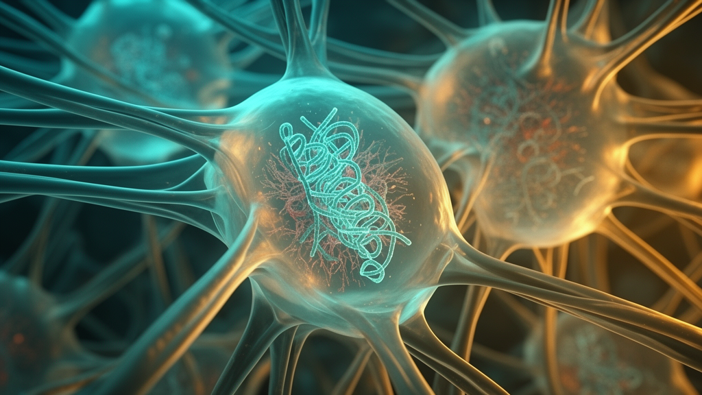
Image Credits: AI Generated
DOI: 10.3389/fcell.2026.1936991
Keywords: lung cancer, cancer cell biology, mechanobiology, tumor microenvironment, genomic instability, oxidative stress, mechanochemical coupling, tissue stiffness, cyclic stretch, NADPH oxidase 2, mitochondrial fragmentation, p120-catenin, epithelial-mesenchymal transition, Nrf2 pathway, mechanopharmacology, chromatin remodeling, DNA repair, lung tumorigenesis, biomechanical landscape, cellular signaling
News Source: Juliet Wilcox. (November 16, 2026). Lung Tissue Tension May Drive Genomic Instability and Malignant Progression. Scienmag.