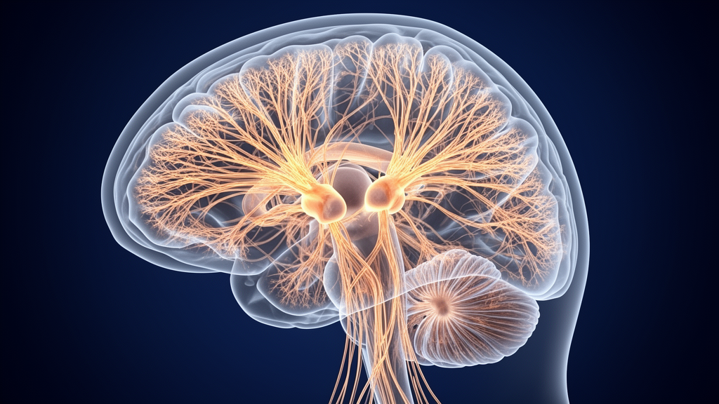
Analyses of magnetic resonance imaging scans from 2,187 patients diagnosed with multiple sclerosis demonstrate that grey matter atrophy spreads outwards from specific cortical regions functioning as disease epicenters. In a peer-reviewed investigation led by Mar Barrantes-Cepas and colleagues, published in the journal Science Advances, researchers found empirical evidence reinforcing the nodal stress hypothesis. This framework posits that the high metabolic strain sustained by heavily active, densely wired regions of the brain gradually exhausts local cellular populations, creating cellular dysfunction that eventually culminates in measurable tissue atrophy.
A recognized hallmark of multiple sclerosis is progressive grey matter atrophy, a structural change that manifests early on magnetic resonance imaging and subsequently disperses throughout the cerebrum in distinct, non-random trajectories. Barrantes-Cepas and the research team explored how these complex anatomical patterns emerge and how they vary between patients across time. By evaluating the extensive dataset of 2,187 participant scans, the authors determined that critical neural hubs possessing greater degrees of connectivity display significantly higher vulnerability to multiple sclerosis atrophy.
The investigators mapped these critical hubs to several prominent cortical and subcortical locations, specifically highlighting the visual cortices, the sensorimotor cortices, and the temporal cortices, as well as the hippocampi, the thalami, and the amygdala. According to the study, these interconnected anatomical structures serve directly as primary disease epicenters because their elevated baseline metabolic demands wear down local brain cells.
In addition to local cell exhaustion within hub regions, the patient scans provided tangible evidence for transneuronal degeneration, the pathological process whereby neurodegeneration established at a primary disease epicenter cascades along structural circuits to affect more distant, connected neurons.
Barrantes-Cepas and coauthors conclude that network-based measures, which can be readily obtained from conventional magnetic resonance imaging and established normative datasets, hold substantial utility for explaining future atrophy progression in individual patients over the course of their disease.
Subject of Research: Brain network connectivity and grey matter atrophy epicenters in multiple sclerosis
Article Title: Busier brain regions can become epicenters for atrophy in multiple sclerosis
Article References: Busier brain regions can become epicenters for atrophy in multiple sclerosis. Original publication (Bibliographic details unavailable.)
Image Credits: AI Generated
DOI: Not provided
Keywords: multiple sclerosis, grey matter atrophy, nodal stress hypothesis, disease epicenters, transneuronal degeneration, neural hubs, metabolic stress, brain connectivity, magnetic resonance imaging, sensorimotor cortex, temporal cortex, visual cortex, hippocampus, thalamus, amygdala, neurodegeneration, Science Advances, Mar Barrantes-Cepas
News Source: Cassandra Pierce. (November 11, 2026). Highly Connected Brain Hubs Act as Epicenters for Multiple Sclerosis Atrophy. Scienmag.