In an in vitro and post-mortem tissue investigation, researchers demonstrated that the ciliary p75 neurotrophin receptor actively facilitates the cellular enrichment of exogenous amyloid beta 1–42 peptide and drives subsequent oxidative stress within human hippocampal astrocytes. Extracellular accumulation of amyloid beta 1–42 peptide represents an established pathological hallmark of Alzheimer’s disease that emerges during the early stages of neurodegeneration. While astrocytes function as resident immune cells that help clear extracellular amyloid peptide from the central nervous system, the precise molecular receptors and cellular organelles governing this uptake have remained incompletely understood. Primary cilia are nonmotile, microtubule-based organelles projecting from astrocytes and neurons that serve as specialized signaling hubs. In this investigation, the research team sought to determine whether astrocytic primary cilia directly engage extracellular amyloid beta through defined membrane receptors, evaluating primary human hippocampal cell cultures alongside post-mortem brain specimens procured from diagnosed Alzheimer’s patients.
To investigate these cellular mechanisms under controlled laboratory conditions, the authors cultured validated primary human hippocampus-derived astrocytes alongside cortex-derived astrocytes for marker validation. Immunofluorescence and flow cytometry confirmed the expression of astrocyte-specific intracellular and transmembrane proteins, including glial fibrillary acidic protein, glutamate-aspartate transporter, and fatty acid binding protein 7. Cortex-derived astrocytes displayed significantly enriched glutamate-aspartate transporter and fatty acid binding protein 7 expression relative to hippocampal astrocytes, matching previously documented baseline tissue profiles. The researchers subsequently treated validated hippocampal astrocytes with graded concentrations of exogenous amyloid beta 1–42 peptide spanning 0.1 to 5 micromolar for durations of 1 hour or 24 hours. To distinguish the experimental treatment from physiologically produced endogenous peptide, the exogenous amyloid beta was conjugated to a HiLyte Fluor 647 fluorochrome label, permitting precise tracking of peptide movement and cellular accumulation across varied exposure times.
Flow cytometric measurements established a rapid, dose-dependent accumulation of exogenous labeled amyloid beta inside human hippocampal astrocytes as early as 1 hour after exposure. At this initial 1-hour time point, treatment with 1 micromolar and 5 micromolar amyloid beta yielded greater than fivefold and tenfold increases, respectively, in total cellular peptide compared to the 0.1 micromolar group. Following 24 hours of exposure, total exogenous amyloid beta content expanded more than tenfold across all evaluated concentrations relative to the 1-hour baseline. To resolve the spatial distribution of the peptide between the plasma membrane and the internal cytoplasm, the researchers applied an anti-amyloid beta antibody under unpermeabilized versus permeabilized membrane conditions. In unpermeabilized cells, where antibody binding is restricted exclusively to surface-bound peptide, membrane-associated amyloid beta rose approximately threefold in the 1 micromolar group and greater than fourfold in the 5 micromolar group at 1 hour, maintaining these high levels through 24 hours.
Permeabilization permitted measurement of total cellular peptide content, allowing the researchers to calculate the relative cytosolic fraction through standardized ratios of permeabilized to unpermeabilized signals. Cytosolic amyloid beta concentrations showed no detectable alteration at 1 hour post-treatment, but increased significantly after 24 hours in the 1 micromolar and 5 micromolar cohorts. These quantitative findings indicated that exogenous amyloid beta 1–42 concentrates predominantly along the outer plasma membrane of hippocampal astrocytes prior to prolonged internalization. The physical accumulation of extracellular peptide directly influenced astrocyte survival and fundamental metabolic health. Utilizing fluorometric quantification of reactive oxygen species, the team recorded an approximate twofold elevation in oxidative stress markers within astrocytes treated with 1 micromolar amyloid beta for 24 hours, with reactive oxygen species rising significantly higher at 5 micromolar concentrations, confirming that sustained surface accumulation triggers pronounced oxidative cellular damage.
Cell viability assays revealed that amyloid beta accumulation drove programmed cell death across the cultured astrocyte populations. Flow cytometric assessment using Annexin V staining indicated that apoptotic markers increased in direct proportion to graded amyloid beta concentrations, mirroring the escalation observed in reactive oxygen species. Median fluorescence intensity of Annexin V climbed significantly even at the lowest concentration of 0.1 micromolar, alongside clear increases in Annexin V-positive cell counts within both live and dead astrocyte fractions. Although Annexin V median fluorescence intensity did not differ significantly between the 1 micromolar and 5 micromolar concentrations, overall apoptotic induction remained robust. Concurrently, cell cycle evaluations combining the proliferation marker Ki67 and violet DNA-selective stains revealed pronounced cycle disruptions at 24 hours. While 0.1 micromolar treatments preserved normal cell cycle distributions, exposures to 1 and 5 micromolar amyloid beta prompted a hyperproliferative Ki67-positive state characterized by expanded G1 and S-phase fractions and significantly contracted G2M fractions.
Investigating the receptor mechanisms underlying this membrane binding, the researchers examined three candidate surface receptors known to interact with amyloid peptides: p75 neurotrophin receptor, CD36, and CD47. Flow cytometry demonstrated striking upregulation of p75 neurotrophin receptor along the astrocyte membrane following amyloid beta treatment. Compared to untreated control cells, surface-associated p75 neurotrophin receptor climbed approximately 20-fold, greater than 100-fold, and roughly 300-fold following 1 hour of exposure to 0.1, 1, and 5 micromolar amyloid beta, respectively, with expression amplifying by an additional tenfold at 24 hours. Cytosolic p75 neurotrophin receptor remained undetectable at 1 hour before demonstrating a transient increase at 24 hours. In contrast, while CD36 and CD47 were detectable on astrocytes, their absolute median fluorescence intensities remained strikingly lower than p75 neurotrophin receptor, and CD47 levels exhibited no significant quantitative changes after amyloid beta application, highlighting the unique reactivity of p75 neurotrophin receptor.
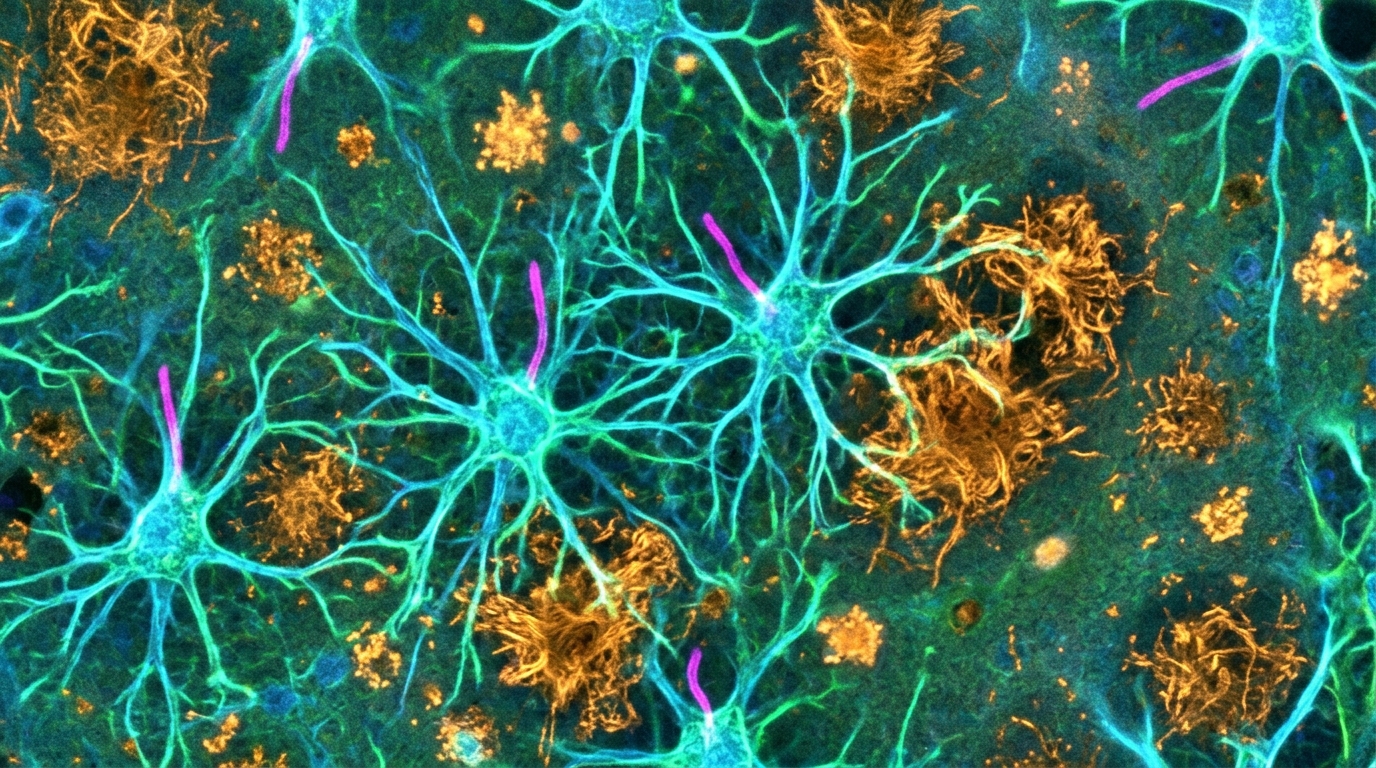
Confocal microscopy confirmed that the upregulated p75 neurotrophin receptor localized prominently to primary cilia, which were labeled using the structural ciliary axoneme marker Arl13b. Exogenous fluorochrome-conjugated amyloid beta 1–42 colocalized directly with ciliary p75 neurotrophin receptor on the cultured astrocytes. Quantitative image analysis demonstrated that the proportion of cilia expressing p75 neurotrophin receptor increased significantly after peptide treatment, with approximately 70 percent of detected exogenous amyloid beta signal colocalizing directly over ciliary p75 neurotrophin receptor complexes. Interestingly, amyloid beta treatment did not significantly alter the overall length or frequency of the astrocytic primary cilia themselves. Subsequent cell cycle profiling established that both exogenous amyloid beta uptake and p75 neurotrophin receptor expression varied across specific division stages, reaching significantly higher levels in G2M-phase astrocytes than in G0-phase astrocytes across all tested concentrations, suggesting stage-dependent modulation of receptor availability and ligand binding.
To evaluate whether these laboratory observations corresponded to human disease states, the authors examined post-mortem hippocampal tissue specimens obtained from Alzheimer’s patients and age- and sex-matched healthy controls. The Alzheimer’s cohort included male donors aged 74 to 86 years (median age 80, n = 4) and female donors aged 57 to 83 years (median age 70, n = 4), while control groups comprised male donors aged 59 to 76 years (median age 67.5, n = 4) and female donors aged 63 to 82 years (median age 72.5, n = 5). Confocal imaging of glial fibrillary acidic protein-positive cells confirmed a higher abundance of astrocytes per field in Alzheimer’s hippocampus compared to controls across both sexes. Astrocytes in Alzheimer’s tissue exhibited significantly higher p75 neurotrophin receptor expression, and nearly the entire receptor signal colocalized with the ciliary marker Arl13b. Although overall astrocyte abundance was comparable between sexes, female Alzheimer’s tissues displayed more pronounced p75 neurotrophin receptor enrichment than male samples.
To establish whether p75 neurotrophin receptor functions as an essential mediator of amyloid beta uptake and downstream oxidative stress, the investigators performed small interfering RNA knockdown experiments. Primary human hippocampal astrocytes were transfected with 5 nanomolar p75 neurotrophin receptor siRNA or non-targeting scramble siRNA for 48 hours prior to 24 hours of exposure to 1 micromolar exogenous amyloid beta. Knockdown produced an approximate 40 percent reduction in plasma membrane-associated p75 neurotrophin receptor without altering cytosolic receptor pools. This targeted membrane receptor depletion caused a corresponding 30 percent reduction in total cellular accumulation of exogenous amyloid beta. Furthermore, while basal reactive oxygen species remained unaffected in the absence of amyloid beta, p75 neurotrophin receptor knockdown produced an approximate 40 percent decrease in cellular reactive oxygen species following peptide exposure, directly coupling receptor presence to amyloid uptake and intracellular oxidative stress generation.
The study provides direct evidence that astrocytic primary cilia express the p75 neurotrophin receptor, which coordinates the capture of extracellular amyloid beta 1–42 and drives pathological oxidative stress responses in human hippocampal astrocytes. By trapping extracellular peptide along their membranes and cilia, astrocytes may initially protect neighboring neurons, but this sequestration triggers sustained reactive oxygen species generation, aberrant S-phase cell cycle delays, and apoptotic astrocyte death. The authors noted that cilia typically disassemble during S-phase to permit cell division, suggesting that amyloid-induced disruptions may impair normal ciliary disassembly dynamics. Important study limitations include the small post-mortem cohort size, which leaves sex-specific expression differences preliminary, alongside unknown genetic backgrounds and clinical disease severities among donor tissues. Nevertheless, the functional coupling of ciliary p75 neurotrophin receptor to amyloid enrichment and oxidative signaling identifies a specific molecular pathway governing astrocyte pathology in Alzheimer’s disease.
Subject of Research: Astrocytic ciliary p75 neurotrophin receptor mechanisms in Alzheimer's disease pathology
Article Title: Ciliary p75 neurotrophin receptor (p75NTR) facilitates the enrichment of exogenous amyloid beta (Aβ 1–42) peptide and promotes oxidative stress in human hippocampal astrocytes
Article References: Gupta, A., Thirugnanam, K., Bice, Z., Lowman, A. K., Rarick, K. R., LaViolette, P. S., Pan, A., Franczak, M., & Ramchandran, R. (2026). Ciliary p75 neurotrophin receptor (p75NTR) facilitates the enrichment of exogenous amyloid beta (Aβ 1–42) peptide and promotes oxidative stress in human hippocampal astrocytes. BMC Molecular and Cell Biology, 27(1), Article 23. https://doi.org/10.1186/s12860-026-00581-z
Image Credits: AI Generated
DOI: 10.1186/s12860-026-00581-z
Keywords: Alzheimer's disease, astrocytes, primary cilia, p75NTR, amyloid beta, neurobiology, oxidative stress, hippocampus, Ciliary, neurotrophin, receptor, facilitates
News Source: Drew Townsend. (November 6, 2026). Astrocytic Cilia Receptor Drives Amyloid Beta Accumulation and Brain Cell Stress. Scienmag.