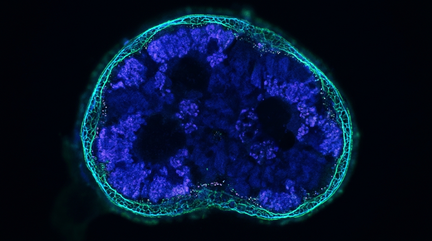
Stem cells age, and one of the clearest molecular fingerprints of that decline is the slow erosion of heterochromatin, the tightly packed, gene-silencing compartment of the genome. A study published in Aging Cell now reports that an enzyme better known for untangling DNA during replication and transcription, topoisomerase II alpha (TOP2A), acts as a guardian of that compartment in human mesenchymal stem cells (MSCs). The researchers found that TOP2A directly binds regulatory sequences of the LMNB1 gene, which encodes the nuclear envelope protein Lamin B1, and that sustaining LMNB1 expression keeps the nuclear membrane and heterochromatin-associated proteins intact. When TOP2A falls, Lamin B1 follows, heterochromatin markers dissipate, and the cells slide into senescence.
The team began with a computational screen rather than a hypothesis. By merging two public RNA-sequencing datasets of MSCs from different donors and passages (GSE35957 and GSE30391, totaling 11 young and 11 senescent samples), they identified 328 upregulated and 586 downregulated genes with an absolute log2 fold change of at least 1. Proliferation markers such as MKI67, PCNA, and MCM2 dropped in the senescent group, while the senescence markers CDKN1A and CDKN2A rose, confirming that the comparison captured genuine senescence. Applying stricter thresholds (absolute log2FC greater than 3 and adjusted p below 0.05) produced a 26-gene senescence set, and weighted gene co-expression network analysis of the same data yielded an 83-gene module that included TOP2A alongside DNA repair and genome stability genes such as RAD51, BARD1, and FANCI. A third gene set, derived from single-cell RNA-sequencing data comparing young (CDKN1A-negative, MKI67-positive) and senescent (CDKN1A-positive, MKI67-negative) MSCs, also contained TOP2A. The intersection of all three sets converged on TOP2A as a core senescence-associated factor.
To test whether that correlation reflects biology, the researchers isolated MSCs from human umbilical cord and passaged them until they became replicatively senescent. Early-passage cells, defined as passage 4, showed the compact morphology expected of healthy MSCs, whereas late-passage cells at passage 13 were enlarged, flattened, and granular, with fragmented nuclei. SA-β-gal staining, a classic marker of senescent cells, was far more abundant in late-passage cultures, and EdU incorporation showed a marked loss of DNA synthesis. Colony-forming ability, a measure of stem cell fitness, also declined significantly. Immunofluorescence and western blotting revealed the reciprocal pattern the screen predicted: p16 protein rose while TOP2A fell in late-passage cells. The downregulation held across MSCs from different donors and across senescent MSCs from different tissues in two additional public datasets, and a multi-organ aging database showed TOP2A expression declining with age in human lung, spleen, bladder, stomach, liver, and intestine.
Correlation alone cannot establish causation, so the team then removed TOP2A experimentally. Transfecting MSCs with TOP2A siRNA induced senescence, and a pharmacological approach confirmed the result: PluriSIn #2, a TOP2A-specific inhibitor that suppresses TOP2A transcription, produced the same phenotype while leaving the related enzyme TOP2B unchanged. Inhibitor-treated MSCs accumulated far more SA-β-gal-positive cells than DMSO controls, formed fewer colonies, and developed enlarged nuclei. Fluorescence intensity of the senescence markers p21 and p16 increased as TOP2A fluorescence decreased, and western blots confirmed the shift at the protein level. The trilineage differentiation capacity of MSCs—their ability to become osteoblasts, adipocytes, and chondrocytes—was impaired after inhibitor treatment, and gene set enrichment analysis of transcriptome data showed upregulation of cellular senescence and senescence-associated secretory phenotype (SASP) pathways alongside downregulation of proliferation pathways. Classical SASP genes, including IL6, IL1A, IL1B, MMP3, AREG, EREG, PAPPA, SERPINE1, and SERPINE2, were all upregulated. Mitochondrial membrane potential dropped, EdU incorporation fell, and flow cytometry showed an increased proportion of cells arrested in G1 phase, consistent with TOP2A’s known cell-cycle dependence.
The mechanistic thread connecting TOP2A to senescence emerged from a trio of genome-wide assays. ATAC-seq mapped chromatin accessibility, CUT&Tag identified the genomic sites actually bound by TOP2A, and RNA-seq quantified the transcriptional consequences. Inhibitor treatment altered 2,134 genes upward and 1,790 downward. CUT&Tag signal concentrated near transcription start sites, and TOP2A-bound sequences were distributed mainly across distal intergenic regions and introns, with a smaller subset at promoters, untranslated regions, and exons, suggesting dual roles in global chromatin architecture and local transcription initiation. Intersecting the three datasets narrowed the field to five candidate genes: MKI67, LMNB1, DMC1, DEPDC1, and CLSPN. ATAC-seq tracks showed reduced chromatin accessibility at the LMNB1 transcription start site, and CUT&Tag followed by quantitative PCR confirmed that TOP2A binds two promoter sequences and two enhancer sequences of LMNB1. A dual luciferase reporter assay in 293T cells overexpressing FLAG-tagged TOP2A strengthened the case: the LMNB1 promoter drove significantly stronger luciferase activity in TOP2A-overexpressing cells than in empty-vector controls, indicating that TOP2A directly activates LMNB1 transcription.
With LMNB1 identified as the direct target, the downstream consequences came into focus. Western blotting and immunofluorescence showed that Lamin B1 and the Lamin B receptor (LBR) were downregulated in inhibitor-treated MSCs. The heterochromatin markers H3K9me3, a repressive histone modification, and the nuclear lamina and heterochromatin proteins LAP2, KAP1, and HP1α all declined as well. Gene set enrichment analysis independently revealed downregulation of heterochromatin formation and organization pathways. Critically, the effect was selective: knockdown of TOP2A significantly reduced Lamin B1, H3K9me3, and KAP1, while Lamin B2, Lamin A/C, and the methyltransferase SUV39H1 were only mildly affected. This specificity pointed to Lamin B1 as the key mediator rather than a general collapse of nuclear lamina proteins.
Rescue experiments sealed the logic. When the researchers overexpressed LMNB1 in MSCs lacking TOP2A, the heterochromatin-associated factors HP1α, KAP1, and H3K9me3 were restored, demonstrating that Lamin B1 functions as a primary downstream effector of TOP2A in maintaining heterochromatin stability. Combined knockdown of both TOP2A and LMNB1 reduced HP1α, KAP1, and H3K9me3 to a magnitude comparable to LMNB1 knockdown alone, further supporting LMNB1 as the critical node. Conversely, overexpressing TOP2A itself in MSCs subjected to serial replicative passaging slowed senescence: TOP2A-overexpressing cells showed less SA-β-gal positivity, better colony formation, higher levels of Lamin B1, LBR, H3K9me3, LAP2, and KAP1, smaller average nuclear size, preserved mitochondrial membrane potential, and greater proliferative activity than empty-vector controls.
To test whether the mechanism extends beyond cultured cells, the team turned to mice. They compared naturally aged C57BL/6J mice at 20 months with young mice at 6 to 8 weeks, and separately treated 10-week-old mice with the TOP2A inhibitor PluriSIn #2 at 5 mg/kg twice weekly for two months. The inhibitor-treated animals developed hair thinning and loss resembling natural aging. SA-β-gal-positive cells increased in the hippocampal region and cortical layers of both old and inhibitor-treated brains. Western blotting of brain tissue showed that TOP2A, Lamin B1, LBR, H3K9me3, LAP2, and KAP1 were all downregulated in naturally aged mice, and that TOP2A, Lamin B1, H3K9me3, and LAP2 declined in inhibitor-treated mice. Behavioral testing revealed reduced locomotor activity in the open field, diminished novel object recognition, and impaired Y-maze spontaneous alternation in both aged and inhibitor-treated animals, although the authors note that the recognition index change in the inhibitor group did not reach statistical significance (p = 0.097) and one Y-maze measure also fell short (p = 0.254), so the behavioral evidence is suggestive rather than uniform.
The authors are careful about what the mouse data do and do not show. They write that the correlative observations in hippocampus and cortex raise the possibility that the TOP2A–LMNB1 axis operates in the brain during aging, but that it remains to be determined whether TOP2A regulates brain aging primarily through this axis or through other TOP2A-dependent mechanisms, such as direct regulation of heterochromatin-related genes or topoisomerase activity-mediated genomic stability. Dissecting these possibilities, they note, will require cell-type-specific manipulation of TOP2A and LMNB1 in the brain. They also flag open questions about upstream regulators of TOP2A and whether the mechanism is conserved in other cell types such as neurons or hepatocytes.
Even with those caveats, the study assembles a coherent chain of evidence in human MSCs: TOP2A expression falls with replicative senescence across donors and tissues; experimental TOP2A loss is sufficient to induce senescence, SASP, mitochondrial dysfunction, and cell-cycle arrest; TOP2A binds LMNB1 promoter and enhancer sequences and activates LMNB1 transcription; Lamin B1 loss destabilizes heterochromatin markers; and restoring either LMNB1 or TOP2A reverses key phenotypes. The work positions TOP2A as a functional link between DNA topology, nuclear architecture, and stem cell aging, and suggests that preserving nuclear scaffold resilience—rather than targeting senescence downstream—might offer a route to maintaining stem cell vitality. Whether that insight can be translated into interventions for human aging remains an open question, but the identification of a single enzyme that simultaneously safeguards genome topology and heterochromatin integrity gives the field a concrete molecular handle to pursue.
Subject of Research: How the topoisomerase TOP2A sustains Lamin B1 and heterochromatin stability to counteract senescence in human mesenchymal stem cells and mouse brain tissue.
Article Title: TOP2A Maintains Heterochromatin Stability by Sustaining LMNB1 Expression to Counteract Senescence
Article References: Cheng, K., Wang, Y., Pan, Y., Zhang, W., Yu, D., Zhou, Q., Zhong, Z., Mei, R., Xu, C., Hayierhan, A., Ma, Y., Xu, Y., Xie, Q., Sun, P., Zhou, J., & Zhou, X. (2026). TOP2A Maintains Heterochromatin Stability by Sustaining LMNB1 Expression to Counteract Senescence. Aging Cell, 25(11), Article e70787. https://doi.org/10.1111/acel.70787
Image Credits: AI Generated
DOI: 10.1111/acel.70787
Keywords: TOP2A, Lamin B1, heterochromatin, mesenchymal stem cells, senescence, nuclear lamina, LMNB1, aging, epigenetics, C57BL/6J mice, SA-β-gal, mitochondrial membrane potential
News Source: Beatrice Stafford. (November 6, 2026). Topoisomerase TOP2A Keeps Stem Cells Young by Guarding Lamin B1 and Heterochromatin. Scienmag.