Veterinary surgeons treating colic in ponies have long worked with a diagnostic blind spot: computed tomography, so useful in dogs and cats, has been largely off-limits for the equine trunk because standard scanners are simply too small. A new anatomical study from Hungary now shows what becomes possible when a large-bore scanner meets a small horse. Researchers at the University of Veterinary Medicine Budapest scanned two Shetland ponies with a 90-centimetre gantry, mapped the normal cross-sectional anatomy of the thorax and abdomen against frozen anatomical sections, and then applied that reference to a live colic case in which the findings shaped the surgical plan.
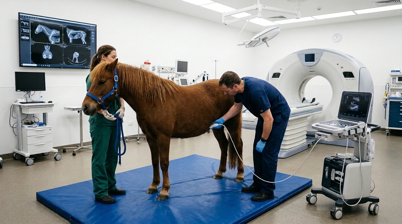
The study, published in Acta Veterinaria Scandinavica, addresses a genuine gap. Whole-body CT atlases exist for species such as houndsharks and dogs, but no comparable reference has been available for the chest and abdominal cavities of horses or ponies. In equine practice, CT has been confined mainly to the head, neck and limbs, because medium-to-large horses cannot fit through a standard gantry. Large-gantry systems change that equation: a 90-centimetre bore can accommodate small-to-medium horses weighing up to about 460 kilograms, which brings ponies and foals comfortably within reach.
The first pony in the study was a 12-year-old mare weighing 130 kilograms, euthanised for welfare reasons after an open metatarsal fracture that the owner declined to treat surgically. According to the owner, the animal had shown no respiratory or colic signs in the previous six months, and no abnormalities were found on clinical examination of the thorax or abdomen. Immediately after euthanasia, the cadaver was scanned in dorsal recumbency with a Canon Toshiba Aquilion Prime SP 16-slice helical multidetector scanner, using 2-millimetre slices, 135 kilovolts and 450 milliamperes. The body was then frozen for three months and cut into 39 transverse slices, each 2 centimetres thick, from the manubrium of the sternum to the rectum, using a band saw. Photographs of the slices, cleaned with ethyl alcohol for sharpness, were matched against the CT images level by level.
Perfect alignment between the two image sets proved impossible, the researchers report. Freezing and post-euthanasia gas production in the intestinal tract shifted volumes slightly, but the discrepancies were minimal and did not hinder identification. Most organs in the anatomical sections were macroscopically visible, and the team annotated 163 structures in the CT images, from the spinal cord, aorta and heart chambers to the liver, spleen, kidneys, adrenal glands, stomach, cecum, the four segments of the large colon, small colon, uterus and ovaries. Major blood vessels were clearly visualised, although much of the fine abdominal vasculature, particularly within the mesocolon, could not be resolved. The right ovary escaped detection, most likely because of its small size and its position outside the sectioning plane.
Some structures resisted even this detailed approach. Muscles, which share similar radiodensity, could only be distinguished on CT where they were bordered by fatty connective tissue; otherwise their boundaries blended together. The small intestines were especially difficult to tell apart, with the descending duodenum and ileum identified largely by position rather than by intrinsic appearance. Nerves and serous membranes, apart from the spinal cord, could not be clearly identified on unenhanced images. Two intestinal positions also deviated from textbook descriptions: the pelvic flexure, filled with contents, sat displaced caudally within the bony pelvis, and the apex of the cecum appeared at the level of the fourth lumbar vertebra on the left side. The authors suggest the flexure may have become impacted during the more than 24 hours the pony spent with a broken limb, and note that no literature establishes whether these positions are normal in Shetland ponies.
The second pony turned the atlas into a clinical tool. This 9-year-old mare, weighing 125 kilograms, presented with suspected large colon impaction containing a large amount of fluid and sand. She had been kept on sandy ground where she fed from the floor, had shown colic signs for more than 24 hours, and had not passed manure during that period. Her abdomen was distended, her heart rate stood at 70 beats per minute, and a laterolateral radiograph confirmed sand in the ventral colon, with sand also suspected in the cecum. With the owner’s consent, the team performed a comprehensive abdominal and thoracic CT examination preoperatively, under total intravenous anaesthesia, in dorsal recumbency with a 70-centimetre field of view.
The scan revealed a distended cecum and an enlarged colon packed with fluid, gas, ingesta of differing attenuation values and sand, with no sign of displacement or torsion. No intestinal segment appeared clearly thick-walled. The team went further, measuring Hounsfield units, the standardised density values that CT assigns relative to water, in 11 intestinal segments and in the liver and right kidney. Gas proved hypoattenuating, fluid hyperattenuating relative to gas but less dense than soft tissue, partially chewed food moderately hyperattenuating with attenuation similar to intestinal fluid, and unchewed fibrous material appeared as rounded, moderately hyperattenuating areas. Sand, which is hyperattenuating, settled ventrally and showed a granular pattern. Gas-fluid levels were visible where gas accumulated in non-dependent regions of the gut.
Marked abdominal distension from the fluid- and gas-filled loops made it difficult to differentiate intestinal segments, although ventrally settled sand helped partially discriminate them. Distinguishing the intestinal wall from fluid-rich content on unenhanced images was described as a genuine limitation of the technique. Adjacent organs were mildly displaced, but no free gas or significant peritoneal fluid was present. Taken together, the findings were compatible with fluid-rich colonic and caecal impaction. The pony went directly from the scanner to the operating room, where surgeons evacuated the impacted cecum and large colon through enterotomies of the cecum and pelvic flexure, exactly as the preoperative CT had anticipated. No thick-walled segments were found at surgery, matching the scan. The intestinal contents were examined macroscopically to confirm gas, fluid, sand and chewed material, and the mare recovered without adverse events, being discharged 10 days after surgery.
The authors are careful about the limits of their work. The study included only two ponies, a small number they explicitly acknowledge as a limitation. The cadaver scan, performed post mortem, cannot reproduce the physiological and motion artefacts of a live anaesthetised horse, and the anatomical deviations in the first pony did not match the literature. Unenhanced CT in large animals has inherent difficulty resolving thin bowel walls, since voxels comparable to wall thickness cause partial volume effects, and contrast CT of equine abdominal organs has not yet been described. The Hounsfield measurements for four content types were confirmed intraoperatively in a single case, and the authors state that these findings require additional confirmation. They also propose that CT cross-sectional imaging may offer more accurate sand detection than radiography, which yields only subjective scoring systems or two-dimensional measurements of a three-dimensional burden, but they flag this as a hypothesis needing further study.
Even with those caveats, the study sketches a practical future for equine trunk imaging. The anatomical reference created from the first pony proved valuable for interpreting the clinical images of the second, and the authors suggest the work could aid not only colic surgery but also the planning of laparoscopic procedures in dorsal recumbency. CT is becoming more accessible in equine clinics worldwide, and thoracic imaging in this case showed no abnormalities, no gravity-dependent atelectasis and no motion artefacts over a 10-minute scan. The authors call for future studies including stallions and geldings to capture pelvic anatomical variation, and for larger case series to confirm the clinical application of their findings. For now, the message is concrete: in ponies and small horses, a large-bore scanner can image the entire trunk, characterise intestinal contents by density, and give surgeons a clearer picture before the first incision.
Subject of Research: Computed tomography anatomy of the pony thorax and abdomen with a clinical colic application
Article Title: Computed tomography anatomy of the pony thorax and abdomen and an example of a clinical application
Article References: Tóth, P., Földes, B., Bódai, E., Pál, Z., & Kapiller, M. (2026). Computed tomography anatomy of the pony thorax and abdomen and an example of a clinical application. Acta Veterinaria Scandinavica, 68(1), Article 48. https://doi.org/10.1186/s13028-026-00891-4
Image Credits: AI Generated
DOI: 10.1186/s13028-026-00891-4
Keywords: computed tomography, pony anatomy, equine colic, thorax, abdomen, Hounsfield units, sand impaction, colic surgery, veterinary imaging, large-gantry CT, anatomical atlas, Shetland pony
News Source: Ophelia Keating. (November 5, 2026). CT Atlas of the Pony Thorax and Abdomen Aids Colic Surgery Planning. Scienmag.