A scaffold infused with eugenol, the clove-derived compound best known for its role in traditional medicine, has shown encouraging laboratory performance as a candidate material for repairing bone defects. In a study published in Biotechnology for Sustainable Materials, researchers at an Indian institution combined eugenol with sodium alginate, chitosan and beta-tricalcium phosphate to create a porous, bioactive scaffold, then evaluated it through computer-based network pharmacology, molecular docking and a battery of physicochemical and cell-based tests. The work, led by Saranya Srinivasan, Ashwathi Vijayalekha and Ashok Kumar Pandurangan, was published on 5 December 2025 as an open-access research article.
The motivation is practical. Bone defects remain a persistent challenge in orthopedics, and the authors note that fractures account for roughly 150,000 hospitalizations annually, while the cost of treating fragility fractures across 27 European Union countries reached about 37 billion dollars in 2010. Hip fractures carry a mortality rate of 15 to 20 percent. Existing approaches, including graft implantation, surgery and medication, still leave the actual rate of regeneration limited, which is why bone tissue engineering with biomaterials has grown into a wide field. An effective scaffold must be biocompatible, non-immunogenic, porous, mechanically stable and degradable at a controlled rate, and natural polymers are attractive because they are less likely to cause rejection than synthetic compounds.
Eugenol itself has prior credentials relevant to bone. Earlier studies cited by the team report that it has anti-inflammatory and antioxidant properties in tissues and cells, that it can inhibit catabolism and reduce apoptosis in chondrocytes by downregulating interleukin-1 beta, and that it counteracted oxidative-stress damage in tendon stem cells when combined with bone marrow mesenchymal stem cells, primarily through upregulation of the Nrf2/HO-1 pathway. Because tendon and bone repair share cellular mechanisms involving oxidative stress modulation and stem cell-mediated remodeling, the researchers reasoned that eugenol’s antioxidant activity could help protect osteogenic cells under oxidative conditions.
To map eugenol’s potential molecular targets in bone disease, the team turned to network pharmacology. They retrieved eugenol’s canonical SMILES from PubChem, assessed its pharmacokinetic profile with SwissADME, which returned a predicted bioavailability score of 0.55 on a 0 to 1 scale, and used the SuperPred database to predict target genes. Comparing those five predicted targets against 16,550 bone-defect-related genes from GeneCards with the InteractiVenn tool yielded 95 overlapping hub genes. CytoHubba in Cytoscape then narrowed the list to a top ten, including KEAP1, NFKB1, NFE2L2, PSMA3, PSMB5, ACE, MME, SLC2A1, EGLN1 and CYP3A4, whose protein-protein interactions were annotated in the STRING database.
Molecular docking in AutoDock then tested how strongly eugenol binds each hub protein. ACE showed the strongest affinity at minus 7.97 kilocalories per mole, followed by NFE2L2 at minus 6.61 and MME at minus 6.46. Moderate binding was seen for KEAP1 at minus 5.67, NFKB1 at minus 5.5 and CYP3A4 at minus 5.17, while SLC2A1, PSMA3, EGLN1 and PSMB5 bound more weakly, with PSMB5 the weakest at minus 3.41. The authors interpret these computational results cautiously, noting that the lower-affinity targets are not direct regulators of osteogenesis but may exert secondary effects on glucose transport, proteasome regulation and hypoxia signalling. Docking scores are predictions, not proof of activity in living tissue, and the study did not test eugenol’s effects on these genes experimentally.
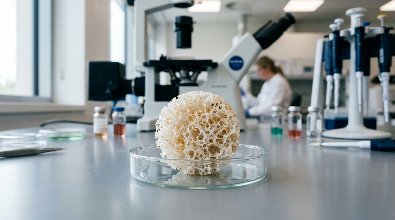
The scaffold itself was built from 1 percent sodium alginate and 2 percent chitosan dissolved in water, with beta-tricalcium phosphate powder added to form a homogeneous suspension. Three groups were prepared: an unloaded control, a version infused with 1 microliter per milliliter of eugenol, and a version with 5 microliters per milliliter. All were crosslinked with 50 millimolar EDC hydrochloride, cast into molds, frozen at minus 80 degrees Celsius and lyophilized to create porous structures. Fourier transform infrared spectroscopy confirmed the functional groups of both polymers and the ceramic, and showed that eugenol’s characteristic peaks were retained with slight shifts in the infused scaffolds, indicating successful incorporation through molecular interactions with the polymeric backbone.
Mechanical testing on a universal testing machine revealed that eugenol improved compressive performance. The control scaffold recorded a peak load of 36 newtons and a compressive strength of 0.073 newtons per square millimeter. Both eugenol-loaded versions reached 50 newtons and 0.102 newtons per square millimeter, with the lower-dose scaffold showing an elongation of 3.923 millimeters and the higher-dose version 5.916 millimeters. The authors attribute the strengthening to hydrogen bonding between eugenol’s hydroxyl group and functional groups in alginate and chitosan, producing a more stable structure rather than a processing artifact. Scanning electron microscopy showed a rough, well-interconnected surface in the eugenol groups, a feature the team links to cell attachment, migration and neovascularization.
Biochemical characterization filled in the rest of the picture. All three groups fell within the 60 to 70 percent porosity range considered optimal for cell infiltration and nutrient diffusion. Swelling behaviour differed by dose: the control and low-dose scaffolds swelled initially and then declined, while the high-dose scaffold showed a continuous increase in swelling over time, which the authors suggest reflects enhanced hydrophilicity and reduced crosslink density at higher eugenol loading. Degradation in lysozyme solution over three weeks was comparable across all groups, indicating that eugenol did not significantly alter the enzymatic degradation rate. Contact angle measurements confirmed hydrophilic surfaces throughout, averaging 57.42 degrees for the control, 89.98 degrees for the low-dose scaffold and 78.04 degrees for the high-dose version, all below the 90-degree threshold for hydrophilicity.
Drug release and biomineralization tests added further detail. Over 24 hours in phosphate-buffered saline, the high-dose scaffold released roughly 68 percent of its eugenol and the low-dose version about 50 percent, both showing an initial burst within the first 8 hours followed by sustained diffusion through the polymeric matrix. After 5 days in simulated body fluid, scanning electron microscopy and energy-dispersive X-ray analysis showed that the low-dose eugenol scaffold developed distinct calcium and phosphorus peaks, suggesting a hydroxyapatite-like mineral layer, while the control surface remained relatively smooth with limited deposition and the high-dose version displayed dense spherical aggregates characteristic of apatite-like crystal growth.
Cell compatibility was assessed with MTT assays on human osteoblast-like MG-63 cells treated with scaffold-conditioned media at 125 micrograms per milliliter. At 24 hours, all conditioned groups showed increased metabolic activity, with the high-dose eugenol scaffold producing the largest significant increase at p below 0.01, and at 48 hours the same group again showed a significant rise at p below 0.001. The authors conclude that the conditioned media is non-toxic and supports osteoblast-like cell proliferation over both short and extended exposure. They caution that future work should include osteogenic gene and protein expression analyses to clarify cellular responses, and that the current findings, being in vitro, do not yet demonstrate bone regeneration in a living system. Still, the combination of computational target identification, mechanical reinforcement, controlled eugenol release and favorable cell response positions this eugenol-infused composite scaffold as a candidate worth pursuing in bone tissue engineering research.
Subject of Research: Development and laboratory evaluation of an eugenol-loaded sodium alginate, chitosan and beta-tricalcium phosphate composite scaffold for bone tissue engineering.
Article Title: Eugenol-loaded sodium Alginate/Chitosan/β-TCP composite scaffold for bone defect regeneration: an in-silico and in-vitro evaluation
Article References: Srinivasan, S., Vijayalekha, A., & Pandurangan, A. K. (2025). Eugenol-loaded sodium Alginate/Chitosan/β-TCP composite scaffold for bone defect regeneration: an in-silico and in-vitro evaluation. Biotechnology for Sustainable Materials, 2(1), Article 25. https://doi.org/10.1186/s44316-025-00048-9
Image Credits: AI Generated
DOI: 10.1186/s44316-025-00048-9
Keywords: eugenol, bone tissue engineering, scaffold, sodium alginate, chitosan, beta-tricalcium phosphate, network pharmacology, molecular docking, MG-63 cells, biomineralization, drug release, biocompatibility
News Source: Gregory Coleman. (November 5, 2026). Eugenol-Infused Alginate-Chitosan Scaffold Shows Promise for Bone Repair in Lab Tests. Scienmag.