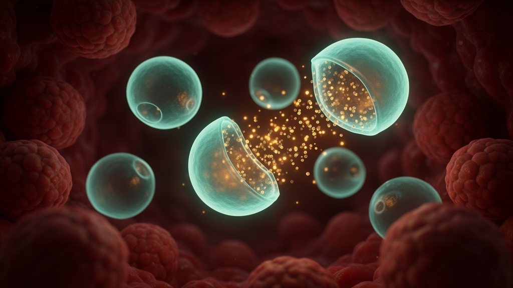
Researchers at Sapienza University of Rome, working with colleagues at the University of Milan, have built a family of pH-sensitive liposomes whose behavior shifts dramatically as the surrounding environment turns acidic, and they have documented that shift with an unusually thorough battery of physical measurements. The study, published in Frontiers in Bioengineering and Biotechnology, describes nanoscale lipid vesicles assembled by microfluidics from the unsaturated phospholipid DOPE, stabilized with oleic acid, and decorated with a cyclic RGD targeting peptide. By tracking particle size, bilayer structure, membrane fluidity and cargo release across a pH ladder from 7.4 down to 4.4, the team showed that the vesicles remain intact at physiological pH but progressively destabilize under the mildly acidic conditions characteristic of late endosomes and lysosomes.
The central design problem the researchers set out to solve is a familiar one in drug delivery. Liposomes are excellent at carrying cargo, but conventional formulations often release it too slowly, and after a cell swallows the vesicle the payload can remain trapped in the endosomal compartment rather than reaching its target. pH-responsive liposomes offer a way around this: they stay stable in the bloodstream at pH 7.4, then fall apart or reorganize when they encounter the acidic interior of endosomes, which sits between roughly pH 4.0 and 5.0, or the acidic extracellular space of tumors, where values between 5.5 and 7.0 are commonly observed. Tumor tissue becomes acidic largely because hypoxic cancer cells rely on anaerobic glycolysis, producing lactic acid, along with excess carbon dioxide generation.
DOPE, or 1,2-dioleoyl-sn-glycero-3-phosphoethanolamine, has long been a favorite building block for such systems because it prefers to pack into structures with negative curvature. On its own, however, it does not readily form stable bilayers, so formulators pair it with helper lipids such as phosphatidylserine, cholesteryl hemisuccinate, or oleic acid. In this study the team chose oleic acid. At neutral pH the deprotonated carboxylate form of oleic acid carries a negative charge, and electrostatic repulsion between these deprotonated groups and the phosphate groups of DOPE helps hold the bilayer together. When the pH drops, the carboxylate becomes protonated, that repulsion weakens, and the membrane is pushed toward destabilization and payload release.
The five formulations the researchers compared increased in complexity step by step. Lipo-DMPC, made of DMPC and cholesterol, served as a non-pH-sensitive control. Lipo-DOPE added DOPE to the DMPC-cholesterol base at a 1:8 molar ratio. Lipo-OA incorporated oleic acid at 5 millimolar alongside 1 millimolar DOPE. The final two formulations, Lipo-cRGD and Lipo-OA-cRGD, added a DOPE-PEG2000-cRGD conjugate, in which the cyclic RGD peptide is tethered to the membrane through a polyethylene glycol spacer. The cRGD motif binds with high affinity to the αvβ3 integrin, a transmembrane receptor involved in cell adhesion, migration and survival signaling that is overexpressed on certain cells, giving the vesicles a targeting handle.
All five formulations were produced by rapid microfluidic mixing rather than conventional bulk preparation. Lipid dissolved in organic solvent and an aqueous buffer were pumped through a Micromixer chip, where chaotic advection and diffusion brought the streams together in milliseconds. The team systematically varied the flow rate ratio and found that a ratio of 20 gave the best compromise between particle size, monodispersity and low multilamellarity, with an aqueous flow of 800 microliters per minute and an organic flow of 40 microliters per minute. After ultracentrifugation to remove residual solvent, the final vesicles measured between roughly 90 and 210 nanometers in hydrodynamic diameter, with polydispersity indices below 0.25. Adding oleic acid shrank the vesicles the most, from about 200 nanometers for the DMPC control down to 89 nanometers, and gave them the most negative surface charge at −42.7 millivolts, a sign of strong electrostatic stabilization.
Small-angle X-ray scattering at the European Synchrotron Radiation Facility in Grenoble provided the structural backbone of the study. At pH 7.4, all formulations showed the scattering signature of vesicular lipid assemblies, with a correlation peak near q = 1 nanometer⁻¹ indicating partially multilamellar organization and an inter-bilayer distance of about 6 nanometers. When the pH was dropped to 5.5, the picture changed sharply for the pH-sensitive formulations. Lipo-DOPE and Lipo-OA both increased in multilamellarity, with the Caillé parameter, a measure of bilayer fluctuation, rising to roughly 0.3 and 0.25 respectively, indicating moderately flexible membrane stacks and a loss of lamellar order. The DMPC control, already highly ordered at physiological pH with a Caillé parameter near 0.1, showed no such change, confirming its role as a non-responsive reference.
Fluorescence probes filled in the dynamic picture. The membrane-embedded probe DPH revealed that anisotropy, which rises as membranes stiffen, fell from 0.28 to 0.14 for Lipo-OA as the pH dropped from 7.4 to 4.4, reflecting enhanced lateral mobility of the lipid chains. Pyrene excimer measurements told a complementary story: Lipo-OA showed the largest increase in excimer emission at acidic pH, consistent with a less tightly packed, more dynamic bilayer, while the PEGylated Lipo-OA-cRGD showed a dampened response, suggesting that the hydrated PEG corona sterically shields the surface and moderates pH-induced rearrangement. Turbidity climbed steeply upon acidification for every formulation except the DMPC control, pointing to pH-dependent aggregation, most pronounced in the oleic acid-containing systems.
Release experiments with two fluorescent cargo molecules, the hydrophilic calcein and the more amphiphilic FITC, quantified the practical consequence of these structural changes. At pH 7.4, Lipo-OA released about 60 percent of its FITC within 48 hours, while Lipo-DOPE held closer to 50 percent over 72 hours. Dropping the pH to 5.5 pushed Lipo-OA to nearly complete FITC release, around 95 to 100 percent after 72 hours, with Lipo-DOPE reaching roughly 75 to 80 percent. At pH 4.4 both approached complete release. Calcein behaved differently: the non-PEGylated formulations leaked substantially even at neutral pH, with Lipo-DOPE releasing about 90 percent of calcein at pH 7.4, a reminder that pH responsiveness does not guarantee cargo retention under physiological conditions.
The cRGD-functionalized, PEGylated formulations preserved the acid-triggered release profile while retaining cargo far better. At pH 7.4 both functionalized formulations released less than about 10 percent of their calcein over 72 hours, and even at pH 4.4 calcein release stayed below roughly 35 percent for Lipo-cRGD and below 50 percent for Lipo-OA-cRGD. FITC release, by contrast, climbed to about 90 percent for the oleic acid-containing formulation at pH 5.5 and nearly complete release at pH 4.4. The researchers attribute the improved calcein retention to the PEG spacer, which forms a steric, hydrated barrier at the vesicle surface that limits water penetration and slows the escape of hydrophilic molecules even when the membrane destabilizes.
Stability testing rounded out the characterization. All formulations kept stable size distributions and surface charges over 90 days of storage at room temperature and at 4 °C, with the refrigerated samples showing slightly less variability. In 45 percent human serum at 37 °C, every formulation showed a transient diameter increase within the first 6 hours, likely reflecting protein corona formation, followed by stabilization through 72 hours, with the cRGD-functionalized systems displaying the most stable size profiles. Serum release studies showed that Lipo-DMPC retained cargo best, while the combined presence of oleic acid and cRGD in Lipo-OA-cRGD produced the fastest release, with nearly complete FITC release within 24 hours. The authors present the work as a proof of concept for pH-responsive, targeted nanocarriers, emphasizing that multi-technique characterization confirmed the pH-responsive behavior survived surface functionalization, and noting that the findings support future development rather than established clinical use.
Subject of Research: pH-sensitive DOPE-based liposomes with oleic acid stabilization and cRGD functionalization for targeted drug delivery
Article Title: From conventional to pH-sensitive formulations: increasing complexity, increasing characterization
Article References: Fabiano, M. G., Giuli, M. V., Stacchini, F. R., Buccini, L., Passeri, D., Del Favero, E., Ricci, C., D’Intino, E., Marianecci, C., & Carafa, M. (2026). From conventional to pH-sensitive formulations: increasing complexity, increasing characterization. Frontiers in Bioengineering and Biotechnology, 14, 1999697. https://doi.org/10.3389/fbioe.2026.1999697
Image Credits: AI Generated
DOI: 10.3389/fbioe.2026.1999697
Keywords: liposomes, pH-sensitive drug delivery, DOPE, oleic acid, cRGD peptide, microfluidics, SAXS, nanocarriers, endosomal pH, PEGylation, fluorescence spectroscopy, tumor microenvironment
News Source: Nathaniel Bowman. (October 28, 2026). Scientists engineer pH-sensitive liposomes that release drugs in acidic tumor environments. Scienmag.