Both were widely used to manage COVID-19 despite preliminary and contradictory findings from clinical trials, and both carry toxic risks when used incorrectly or at excessive doses. A new experimental study published in the Egyptian Journal of Forensic Sciences has now examined in detail what repeated doses of these drugs do to the brain, heart and liver of laboratory rats, and whether vitamin C can soften the blow. The answer, according to the researchers, is that the drugs inflict measurable oxidative, inflammatory and apoptotic damage across all three organs, and that vitamin C co-administration substantially reverses those changes.
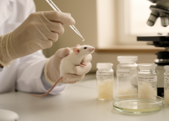
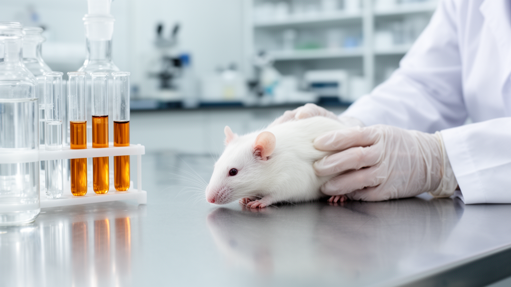
The research team, led by Mennatallah Mahmoud Ahmed Mohamed of the Forensic Medicine and Clinical Toxicology department at their university’s Faculty of Medicine, conducted the laboratory work between October and November 2022. Sixty adult Wistar albino rats, twelve weeks old and weighing roughly 220 to 250 grams, were divided into six groups of ten animals each: a control group, a vitamin C-only group, an ivermectin-treated group, a hydroxychloroquine-treated group, and two groups receiving one of the drugs together with vitamin C. All drugs were given orally through a gastric tube for three weeks, after which the animals were observed for an additional week to detect any delayed toxic effects before being humanely euthanized. The doses were chosen to approximate human equivalent dosing: ivermectin at 3.7 milligrams per kilogram once daily, hydroxychloroquine at 35 milligrams per kilogram twice on the first day followed by 17.5 milligrams per kilogram twice daily, and vitamin C at 176 milligrams per kilogram once daily.
The investigators then subjected the cerebral cortex, heart and liver to a battery of biochemical, histological and immunohistochemical analyses. Serum biomarkers of liver and heart injury, including alanine aminotransferase, aspartate aminotransferase, lactate dehydrogenase and creatine kinase-myocardial band, were measured alongside tissue levels of the oxidants malondialdehyde and nitric oxide and the antioxidants glutathione and superoxide dismutase. Tissue sections were stained with hematoxylin and eosin and Masson’s trichrome to assess structural damage and collagen deposition, and immunostaining for cleaved caspase-3, a widely used marker of apoptosis, quantified the extent of programmed cell death in each organ.
The biochemical results painted a consistent picture of oxidative stress. In the brain, both drugs significantly elevated malondialdehyde and nitric oxide while lowering glutathione and superoxide dismutase compared with control and vitamin C-only animals, with ivermectin producing a stronger oxidant shift and a deeper antioxidant depletion than hydroxychloroquine. In the heart, malondialdehyde rose significantly and creatine kinase-myocardial band, a clinical marker of cardiac muscle injury, climbed sharply in both drug-treated groups. In the liver, the pattern was similar: malondialdehyde increased significantly, antioxidant defenses fell, and the liver enzymes ALT, AST and LDH rose significantly, with hydroxychloroquine producing a particularly pronounced increase in AST and LDH. Interestingly, in cardiac tissue the antioxidants superoxide dismutase and glutathione showed a non-significant increase rather than a decrease in the drug-treated groups, a pattern the authors interpret as a possible compensatory response to oxidative stress amplification.
The histological findings mirrored the biochemical ones. In the cerebral cortex, ivermectin-treated rats showed degenerated granular and pyramidal neurons with pericellular halos, intensely stained pyknotic nuclei, vacuolated neuropil, vascular congestion and hemorrhage beneath the pia mater, while hydroxychloroquine produced multiple degenerated pyramidal neurons and vacuolated neuropil with most granular neurons spared. Morphometric counting confirmed a significant increase in the number of degenerated neurons in both drug groups, with ivermectin again the more damaging of the two. In the heart, ivermectin caused marked distortion of myocytes with hypereosinophilic areas, myofibril separation, corrugation, nuclear pyknosis, widened interstitial spaces, inflammatory cell infiltration and capillary congestion, whereas hydroxychloroquine produced focal degeneration of some myocytes with congested capillaries and inflammatory infiltrates. Liver sections from both drug groups displayed disrupted hepatic architecture, hepatocyte vacuolation, nuclear pyknosis, sinusoidal congestion and dilatation, portal vein dilatation and inflammatory cell infiltration, with hemorrhage around central veins and in the periportal area in the ivermectin group.
Immunohistochemistry for cleaved caspase-3 added an apoptotic dimension to the toxicity picture. Control and vitamin C-only animals showed negative or minimal immune reactivity in all three organs, while both drug-treated groups displayed significantly increased numbers of cleaved caspase-3 positive neurons, myocytes and hepatocytes, along with elevated optical density of the immune reaction. Masson’s trichrome staining revealed significantly increased collagen deposition between myocytes and around central veins and portal areas in the drug-treated hearts and livers, indicating early fibrotic change. In the heart, ivermectin drove collagen deposition to the point of replacing degenerated myocytes, while hydroxychloroquine produced milder but still significantly elevated collagen compared with controls.
The most striking results emerged when vitamin C was co-administered. In the brain, oxidant levels fell significantly and antioxidant levels rose in the combined-treatment groups, with superoxide dismutase returning to control levels and glutathione approaching it. The number of degenerated neurons dropped significantly, and cleaved caspase-3 reactivity declined markedly, with the hydroxychloroquine plus vitamin C group showing a greater reduction than the ivermectin combination group. In the heart, creatine kinase-myocardial band fell back to control values, cardiac oxidants dropped significantly, and collagen deposition was significantly reduced. In the liver, ALT, AST and LDH values fell significantly, though they did not fully return to control levels, and hepatic oxidant markers declined while antioxidants rose. Histologically, vitamin C co-treatment preserved most neurons, myocytes and hepatocytes, with only scattered degenerated cells and minimal residual vacuolation or congestion visible under the microscope.
The authors attribute the protective effect of vitamin C to its role as a free radical scavenger that donates electrons to neutralize reactive oxygen species before they damage lipids, proteins and DNA. Citing prior preclinical work, they note that ascorbic acid also supports glutathione peroxidase function, activates the Nrf2/Keap1 signaling cascade that upregulates antioxidant enzymes such as superoxide dismutase and heme oxygenase-1, and modulates inflammatory and autophagy-related proteins. In the discussion, the team connects their findings to earlier research showing that ivermectin triggers the mitochondrial apoptotic cascade through caspase-3 activation, that hydroxychloroquine toxicity involves oxidative overload and autophagy suppression in cardiomyocytes, and that vitamin C has previously been shown to mitigate ivermectin-induced hepatic and cerebral damage in rabbits and rats.
The study has clear limitations that the authors themselves acknowledge. The findings come from a rat model with species-specific organ physiology and drug pharmacokinetics that may differ substantially from humans, and the dosing period was limited to three weeks, whereas some human patients require these drugs for much longer durations. The authors also note that additional clinical trials are necessary before any application to human practice, and that further studies on other organs and mechanisms are recommended. The authors recommend that ivermectin or hydroxychloroquine be used cautiously and that co-administration of vitamin C is highly recommended, while suggesting that future preclinical research should validate vitamin C as an adjuvant antidote during long-term hydroxychloroquine or ivermectin therapies. No deaths occurred in any group during the experiment, and all animals ate and drank normally throughout.
For now, the study stands as a detailed preclinical demonstration that repeated oral dosing of ivermectin or hydroxychloroquine at human-equivalent doses generates oxidative stress, inflammation, apoptosis and early fibrosis in the rat cerebral cortex, heart and liver, and that concurrent vitamin C administration at 176 milligrams per kilogram per day largely reverses these changes across all measured parameters. Whether the same protective effect holds in humans, at what dose, and over what duration remain open questions that only properly designed clinical trials can answer.
Subject of Research: Toxic effects of ivermectin and hydroxychloroquine on rat brain, heart and liver and the protective role of vitamin C
Article Title: The neurological, cardiac, and hepatic toxic effects of ivermectin and hydroxychloroquine and the ameliorative role of vitamin C: a biochemical, histological, and immunohistochemical experimental study
Article References: Mohamed, M. M. A., Abdelaleem, M. A., Mokhemer, S. A., Albadawi, E. A., Hassan, S. M., Ali, E. A. E., & Asem, M. M. (2026). The neurological, cardiac, and hepatic toxic effects of ivermectin and hydroxychloroquine and the ameliorative role of vitamin C: a biochemical, histological, and immunohistochemical experimental study. Egyptian Journal of Forensic Sciences, 16(1), Article 88. https://doi.org/10.1186/s41935-026-00586-x
Image Credits: AI Generated
DOI: 10.1186/s41935-026-00586-x
Keywords: ivermectin, hydroxychloroquine, vitamin C, oxidative stress, rat study, hepatotoxicity, neurotoxicity, cardiotoxicity, apoptosis, antioxidants, toxicology, Wistar rats
News Source: Ophelia Keating. (October 28, 2026). Vitamin C Shields Rat Brains, Hearts and Livers from Ivermectin and Hydroxychloroquine Damage in Experimental Study. Scienmag.