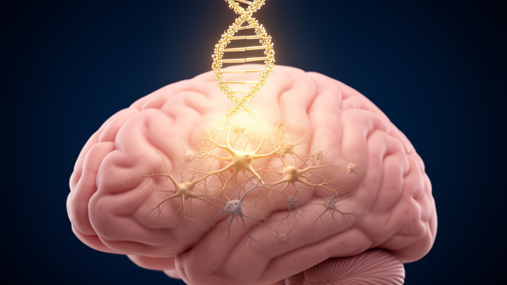
A molecular chain reaction that destroys neurons after birth-related oxygen deprivation may have a weak link, and a new study points to a small RNA molecule as the way to exploit it. In a paper published in iScience, researchers report that a microRNA called miR-7a-5p protects the neonatal brain in experimental hypoxic-ischemic encephalopathy by reining in PARP1, a DNA repair enzyme whose overactivation drives a form of programmed cell death known as parthanatos. The work, conducted in rat models and in a mouse neuronal cell line, positions the miR-7a-5p/PARP1 axis as a candidate therapeutic target for a condition that currently has only one established clinical treatment.
Hypoxic-ischemic encephalopathy arises when a newborn’s brain is starved of oxygen and blood flow around the time of birth. Globally, it affects an estimated 1 to 8 per 1,000 live births and is a major cause of neonatal mortality and long-term neurodevelopmental impairment. Roughly 15 percent of affected newborns die within the first week of life, and about 40 percent of survivors live with permanent neurological sequelae, including cerebral palsy, cognitive impairment, sensory and behavioral disorders, and epilepsy. Therapeutic hypothermia remains the only effective treatment in clinical practice, yet approximately half of infants who receive it still die or sustain permanent neurobehavioral and cognitive dysfunction, leaving an urgent need for adjunctive strategies.
The research team began with a bioinformatics lead rather than a hypothesis about cell death. Mining the publicly available GEO dataset GSE121178, which contains microarray profiles of whole blood from three infants with HIE and three healthy controls, they identified 40 differentially expressed microRNAs using thresholds of an absolute log2 fold change greater than 1 and a p value below 0.05. Among these, the precursor gene miR-7-2 stood out as significantly downregulated in the HIE samples. After processing, miR-7-2 gives rise to the mature miR-7a-5p, a microRNA previously implicated in hypoxic-ischemic brain injury, detected at altered levels in the blood of patients with acute large vessel occlusion stroke, and found at reduced abundance in the brains of stroke rats. Despite these clues, its functional role in HIE had never been characterized.
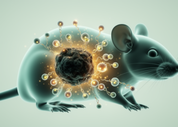
To test function, the researchers used a neonatal rat model of hypoxic-ischemic injury. Injured animals showed clear motor deficits: from postnatal day 9 to 11, model-group rats took longer to right themselves than controls, could hold stable on an inclined plane only at shallower angles, needed more time to cross a balance beam, and slipped more often with their hind limbs while crossing. Real-time PCR then confirmed that miR-7a-5p was downregulated both in the injured cortex and in peripheral blood of the HIE rats, mirroring the human blood signature that first drew the team’s attention.
The mechanistic core of the study concerns PARP1, poly (ADP-ribose) polymerase-1. Using the TargetScan database, the team predicted that miR-7a-5p binds to nucleotide positions 628 to 635 within the 3′ untranslated region of PARP1 messenger RNA. A luciferase reporter assay in HEK293T cells validated the interaction: a miR-7a-5p mimic suppressed the activity of the wild-type PARP1 reporter but had no effect on a mutated version, confirming sequence-specific binding. In the injured cortex of HIE rats, western blotting showed PARP1 hyperactivation, along with elevated levels of PAR polymer and AIF, apoptosis-inducing factor, the downstream effectors of parthanatos. Notably, cleaved-PARP1, a marker of apoptosis, showed no difference between HIE and control tissue, and the authors report that classical apoptosis markers were absent. Electron microscopy revealed fewer mitochondria within cortical neurons, which appeared swollen with disrupted cristae, a pattern consistent with parthanatos-driven damage.
Parthanatos is a caspase-independent, PARP1-mediated form of programmed necrosis first linked to cerebral ischemic injury as early as 1997. When PARP1 becomes hyperactive in response to massive DNA damage, it consumes NAD+ at an unsustainable rate, depleting cellular energy stores and triggering the release of AIF from mitochondria. AIF, together with macrophage migration inhibitory factor, translocates to the nucleus, where it drives extensive DNA fragmentation and chromatin condensation, culminating in rapid necrotic death. The authors argue that their biochemical signature, PAR accumulation and AIF translocation without apoptosis markers, establishes parthanatos as the dominant neuronal death pathway in their experimental HIE model, extending a paradigm previously studied mainly in adult stroke to the neonatal setting.
Therapeutic proof of concept came from an agomir, a chemically stabilized miRNA analogue. Injected intracerebroventricularly six hours after hypoxia-ischemia, the miR-7a-5p agomir significantly reduced cerebral infarct volume measured by TTC staining at 48 hours, compared with rats receiving a negative control oligonucleotide. Electron microscopy showed less mitochondrial damage and chromatin condensation in cortical neurons, and in a novel object recognition test, agomir-treated rats spent a significantly higher percentage of time exploring a novel object than injured controls given the negative control, indicating partial recovery of recognition memory. These experiments used small animal groups, with infarct imaging based on three animals per group and behavioral testing on six, so the results should be read as early preclinical signals rather than established treatment effects.
In vitro, the team used L-glutamate to induce oxidative stress in HT22 cells, a well-established model of excitotoxic neuronal injury in which glutamate blocks the cystine/glutamate antiporter, depletes glutathione, and provokes reactive oxygen species production. Twelve hours of 5 millimolar L-glutamate effectively induced cell death, lowered mitochondrial membrane potential, raised ROS levels, increased PARP1, PAR, and AIF abundance, and drove AIF and MIF into the nuclei. The authors checked whether other cell death programs were running in parallel: ferroptosis markers GPX4 and ACSL4 and necroptosis markers p-RIP3 and p-MLKL showed no significant changes, either in cells or in HIE rat cortex. Pharmacological inhibition of PARP1 with PJ34, which peaked in protective effect at 320 micromolar, attenuated cell death, reduced ROS, eased excessive ATP consumption, and reversed PAR formation and AIF nuclear translocation, underscoring PARP1’s central role in the injury cascade.
Two further experiments tightened the causal chain. Overexpressing miR-7a-5p via lentiviral transfection protected HT22 cells from glutamate: viability recovered, cell death fell, mitochondrial membrane potential and ATP levels were preserved, and PARP1, AIF, PAR, and AIF nuclear localization all decreased. Conversely, activating PARP1 with the agonist MNNG reversed the protection. A 12-hour incubation with 5 micromolar MNNG significantly abolished the viability benefit of miR-7a-5p overexpression, and concentrations of 1 and 5 micromolar reversed its protection against cytotoxicity measured by LDH release. Together, these results indicate that miR-7a-5p suppresses parthanatos in a PARP1-dependent manner.
The authors are candid about the limits of the work. The glutamate-treated HT22 model cannot reproduce the full constellation of HIE pathology, which also involves energy failure, acidosis, neuroinflammation, and blood-brain barrier disruption, and the study did not examine whether the miR-7a-5p/PARP1 axis operates in glial cells, which also respond to miR-7 in other brain injury contexts. No imaging data were collected, and the authors plan functional MRI in future work. Translation faces further hurdles, including delivering miRNA therapeutics across the neonatal blood-brain barrier, defining a therapeutic window, and establishing long-term safety in developing organisms. Still, the authors suggest that strategies to boost miR-7a-5p activity, whether synthetic mimics or agents that enhance endogenous expression, could complement hypothermia by directly targeting parthanatos, while the persistent elevation of miR-7a-5p detectable in the peripheral blood of HIE rats hints at a possible biomarker role. For now, the finding stands as a preclinical demonstration in rats and a neuronal cell line that a single microRNA can dial down a catastrophic cell death pathway in the oxygen-starved newborn brain.
Subject of Research: The role of the miR-7a-5p/PARP1 axis in parthanatos-mediated neuronal death in neonatal hypoxic-ischemic encephalopathy
Article Title: miR-7a-5p attenuates hypoxic-ischemic encephalopathy by suppressing PARP1-mediated parthanatos
Article References: Chen, D., Kong, X., Zhong, H., Zhang, Y., Si, C., Kasimu, A., Qiao, R., Song, Y., & Li, B. (2026). miR-7a-5p attenuates hypoxic-ischemic encephalopathy by suppressing PARP1-mediated parthanatos. iScience, 29(11), Article 117947. https://doi.org/10.1016/j.isci.2026.117947
Image Credits: AI Generated
DOI: 10.1016/j.isci.2026.117947
Keywords: hypoxic-ischemic encephalopathy, miR-7a-5p, PARP1, parthanatos, neonatal brain injury, microRNA, apoptosis-inducing factor, therapeutic hypothermia, HT22 cells, neuroprotection, oxidative stress, preclinical study
News Source: Harold Sullivan. (October 27, 2026). Tiny RNA Molecule Shields Newborn Brains from Oxygen-Starved Cell Death. Scienmag.