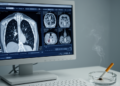
PSMA-PET/CT detected 91% of clinically significant prostate cancers in a retrospective analysis of patients with elevated PSA levels and suspected disease, according to a Medical University of Vienna study. The analysis also found that a normal scan identified a group in which tissue examination confirmed no clinically significant tumour in 91% of cases, suggesting the imaging method could help determine which patients need a biopsy. The findings do not yet establish whether PSMA-PET/CT should replace biopsy or be used routinely in initial prostate-cancer diagnosis.
The study was a post-hoc analysis of the randomised, controlled, phase 3 RAPID trial. It included 194 patients whose elevated blood PSA levels had prompted suspicion of prostate cancer but who had not yet undergone biopsy. Researchers assessed PSMA-PET/CT before tissue sampling and compared its findings with the pathology results. Among 77 patients in whom biopsy confirmed clinically significant prostate cancer, 70 had been identified by the imaging scan. These observational results were obtained within a randomised trial, but the reported imaging analysis itself was retrospective.
PSMA-PET/CT combines positron emission tomography with computed tomography. For the PET component, patients receive a small amount of a radioactively labelled substance that binds to prostate-specific membrane antigen, or PSMA, a protein described as particularly abundant on prostate-cancer cells. Areas that bind the tracer can then be visualised, while the simultaneous CT scan supplies anatomical images that locate suspicious findings within the body’s structures. The method therefore supplies both molecular and anatomical information before a biopsy is performed, although the analysis does not show that using it in routine care would improve clinical outcomes.
The researchers also examined what happened after the initial assessment. During a median follow-up of 35 months, 23 of the 24 patients who subsequently developed an aggressive course of the disease had already been identified by PSMA-PET/CT. This finding indicates that the scan was associated with the detection of many cases that later followed an aggressive course. It does not, by itself, demonstrate that scanning prevented advanced disease or that it can predict the course of an individual cancer more accurately than every alternative diagnostic strategy.
The central potential benefit concerns the interpretation of a normal scan. In the study, 91% of patients with normal PSMA-PET/CT results did not have a clinically significant tumour on tissue examination. That high negative predictive value suggests that pre-biopsy imaging could help clinicians distinguish patients whose suspicion of clinically significant cancer is low from those for whom tissue sampling remains warranted. The result is especially relevant because PSA elevation can indicate prostate cancer but cannot provide a definitive diagnosis, and suspicion commonly leads to a prostate biopsy.
Study lead Marcus Hacker, head of the Division of Nuclear Medicine at the Department of Biomedical Imaging and Image-guided Therapy, said the findings show that PSMA-PET/CT may provide valuable information early in the diagnostic work-up. He particularly highlighted the diagnostic value of a normal result, which could in future help guide decisions about which patients require a biopsy. The team’s conclusion is prospective: further studies are needed to determine whether and how the imaging method should be integrated into routine diagnostics.
The analysis has several limits that constrain stronger conclusions. It was a post-hoc analysis rather than a prospective test of a biopsy-avoidance strategy, and only 194 patients with elevated PSA and suspected prostate cancer were included. Although the study came from a randomised phase 3 trial, the reported role of PSMA-PET/CT was evaluated retrospectively within that setting. The findings therefore support further investigation of pre-biopsy PSMA-PET/CT but do not establish that a normal scan can safely eliminate biopsy, that the method improves long-term outcomes, or that its performance will be identical in broader populations.
Subject of Research: Pre-biopsy PSMA-PET/CT for diagnosing clinically significant prostate cancer in patients with elevated PSA
Article Title: Prostate cancer: Specialized imaging could prevent unnecessary biopsies
Article References: Prostate cancer: Specialized imaging could prevent unnecessary biopsies. Original publication (Bibliographic details unavailable.)
Image Credits: AI Generated
DOI: Not provided
Keywords: prostate cancer, PSMA-PET/CT, PSA testing, prostate biopsy, medical imaging, cancer diagnosis, RAPID trial, negative predictive value, Prostate, cancer, Specialized, imaging
News Source: Nathaniel Bowman. (October 26, 2026). PSMA-PET/CT detected 91% of clinically significant prostate cancers in biopsy study. Scienmag.