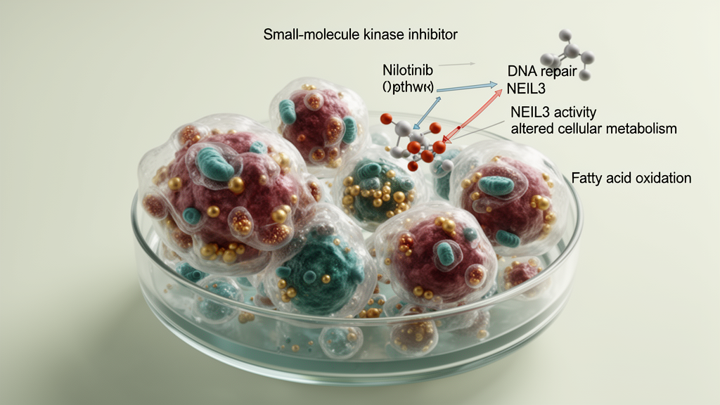
A team of researchers in China reports that the leukemia drug nilotinib can strip hepatocellular carcinoma cells of a key survival mechanism by binding directly to the DNA repair protein NEIL3 and shutting down fatty acid oxidation, a metabolic pathway the cancer cells rely on to resist anoikis, the programmed cell death triggered when cells detach from their surroundings. The study, published in Frontiers in Oncology, combines bioinformatics, cell culture, molecular docking, and mouse xenograft experiments to build what the authors describe as the first evidence for a NEIL3–fatty acid oxidation–anoikis resistance axis in liver cancer, and it positions an already approved tyrosine kinase inhibitor as a candidate for repurposing against one of the world’s deadliest malignancies.
The clinical stakes are considerable. Hepatocellular carcinoma is the sixth most frequently diagnosed cancer worldwide and the fourth leading cause of cancer death, arising against a backdrop of chronic hepatitis, cirrhosis, and a complex interplay of genetic and epigenetic alterations. Although local ablation, surgical resection, and transplantation can be curative in early disease, prognosis remains unfavorable because recurrence and metastasis are frequent. Metastasis begins with a deceptively simple problem: a tumor cell must detach from the extracellular matrix, survive in the circulation, and colonize distant tissue. Anoikis is the programmed cell death mechanism that normally kills detached cells, so cancer cells that evade it gain a decisive survival advantage. Understanding how hepatocellular carcinoma cells overcome anoikis has therefore become a central question in the search for new therapeutic targets.
The study’s starting point was the NEIL3 gene, which encodes a DNA glycosylase of the NEIL family homologous to bacterial Fpg/Nei proteins. NEIL3 initiates DNA base excision repair, modulates lipid metabolism, and has been implicated in cerebral ischemia and autoimmunity, but it also displays clear oncogenic behavior. Previous work showed that in hepatocellular carcinoma NEIL3 activates the BRAF/MEK/ERK/TWIST pathway to promote invasion, migration, and stemness, while in esophageal cancer NEIL3 knockdown promotes apoptosis and delays tumor growth, and in lung adenocarcinoma high NEIL3 expression attenuates DNA damage and fosters cisplatin resistance. What remained unknown was whether NEIL3 influences anoikis in liver cancer.
To address that question, the researchers first mapped NEIL3 expression. Data from The Cancer Genome Atlas, comparing 374 hepatocellular carcinoma tissues with 50 adjacent normal tissues, showed significantly higher NEIL3 mRNA in tumors, a finding the team confirmed by RT-qPCR in ten pairs of patient tumor and matched adjacent non-tumor liver samples collected at The Central Hospital of Yongzhou from patients who had received no prior radiotherapy, chemotherapy, or targeted therapy. NEIL3 was also overexpressed in multiple hepatocellular carcinoma cell lines, including SNU-475, Hep3B, and Huh-7, relative to normal human liver epithelial THLE-3 cells. Critically, survival analysis of the TCGA-LIHC cohort using the GEPIA2 database showed that patients in the high NEIL3 expression group had significantly shorter overall survival than those in the low expression group.
The functional experiments then established NEIL3 as a suppressor of anoikis. Using short hairpin RNA lentiviral particles to stably knock down NEIL3 in Huh-7 cells, the researchers observed reduced proliferation in CCK-8 assays and, under suspension culture on poly-HEMA-coated plates that force cells to detach, increased anoikis by three independent measures: live/dead staining showed enhanced red fluorescence indicating more dead cells, Annexin V-FITC/PI flow cytometry confirmed a significant rise in apoptosis, and Western blotting detected upregulation of the apoptosis markers cleaved-PARP and cleaved-caspase3. Together, these results demonstrated that NEIL3 overexpression in hepatocellular carcinoma promotes cancer cell survival by suppressing anoikis.
The mechanism emerged from gene set enrichment analysis, which revealed that NEIL3 expression was enriched in fatty acid metabolism and oxidation pathways. Pearson correlation analysis showed that NEIL3 was significantly positively correlated with four key fatty acid oxidation genes spanning distinct pathway nodes: PPT1 in substrate hydrolysis, ACSL4 and ACSL3 in cytosolic acyl-CoA activation, and ABCD1 in peroxisomal transport. When the team overexpressed NEIL3 in SNU-475 cells, oxygen consumption rates, ATP levels, and acetyl-CoA levels all rose while free fatty acid content fell, a metabolic signature of enhanced fatty acid oxidation. NEIL3 overexpression also increased cell viability, reduced the proportion of dead cells in live/dead staining, and decreased apoptosis under detachment conditions. All of these effects were reversed by etomoxir, a fatty acid oxidation inhibitor, which restored viability and apoptosis levels to those of control cells, directly supporting the conclusion that NEIL3 suppresses anoikis by activating fatty acid oxidation.
With the target mechanism in hand, the researchers screened for small molecules capable of binding NEIL3 and blocking its function. Batch molecular docking against the NEIL3 protein structure, obtained from the RCSB PDB database under identifier 7JL5 and screened against the DrugBank ligand library using AutoDock Vina, identified nilotinib as the top candidate with the lowest binding free energy. Interaction analysis showed that nilotinib forms hydrophobic contacts with NEIL3 residues Arg175, Asp183, Gln184, and Asn192, and hydrogen bonds with Lys81 and Arg271. Two orthogonal assays validated the binding in Huh-7 cells: in the cellular thermal shift assay, nilotinib treatment increased NEIL3 protein stability upon thermal challenge at graded temperatures from 45 to 80 degrees Celsius compared with the DMSO control, and in the drug affinity responsive target stability assay, nilotinib protected NEIL3 from Pronase E digestion in a concentration-dependent manner.
Functional testing confirmed that nilotinib antagonizes NEIL3’s metabolic and anti-anoikis effects. In SNU-475 cells divided into four groups, nilotinib significantly decreased oxygen consumption rates and ATP levels compared with vehicle control, and NEIL3 overexpression partially counteracted that metabolic inhibition. The CCK-8 assay showed that nilotinib blocked the pro-viability effect of NEIL3 overexpression, while live/dead staining and flow cytometry demonstrated that nilotinib treatment reversed the suppression of anoikis by NEIL3 overexpression. In other words, nilotinib promotes anoikis in hepatocellular carcinoma cells by engaging NEIL3 and counteracting NEIL3-mediated activation of fatty acid oxidation.
The in vivo evidence came from a xenograft model in which twenty-four female BALB/c nude mice bearing subcutaneous tumors of SNU-475 cells stably overexpressing NEIL3 or control vector were randomly allocated to four groups of six and treated with 20 mg/kg nilotinib or PBS by oral gavage every two days. NEIL3 overexpression alone significantly increased tumor volume and weight, but nilotinib markedly suppressed that NEIL3-driven growth over the 25-day treatment period. Immunohistochemistry of harvested tumors showed that NEIL3 overexpression raised Ki67, a proliferation marker, and lowered cleaved-caspase3, while nilotinib treatment reduced Ki67 and increased cleaved-caspase3. Expression of CPT1A, the rate-limiting enzyme of fatty acid oxidation, was significantly higher in NEIL3-overexpressing tumors and reduced by nilotinib. Notably, nilotinib did not alter NEIL3 protein expression itself, indicating that the drug suppresses fatty acid oxidation by binding to NEIL3 rather than by degrading it.
The authors place their findings within a broader survival paradigm: activation of fatty acid oxidation is a common strategy by which tumor cells resist anoikis, because detached cells typically suffer suppressed glucose uptake and catabolism, depleting ATP and NADPH, and fatty acid oxidation restores energy homeostasis. CPT1A-mediated fatty acid oxidation has been shown to confer anoikis resistance in colorectal cancer by scavenging reactive oxygen species, in ovarian cancer, and in esophageal squamous cell carcinoma under detachment conditions. The authors propose that NEIL3, by efficiently repairing oxidative DNA damage and alleviating oxidative stress, may relieve oxidative inhibition of PPARα, the transcription factor that regulates fatty acid oxidation genes including CPT1A, although they caution that whether NEIL3 acts directly as a transcriptional co-regulator at fatty acid oxidation gene regulatory regions or indirectly through its base excision repair activity remains to be elucidated, with chromatin immunoprecipitation sequencing and ATAC-seq proposed as future approaches. Nilotinib itself is a second-generation BCR-ABL tyrosine kinase inhibitor approved for chronic myeloid leukemia, with reported activity in melanoma, colorectal cancer, and hepatocellular carcinoma, and an ability to inhibit ABC transporters such as ABCC10/MRP7 to overcome multidrug resistance. The study’s limitations are substantial: the evidence derives from cell lines, ten paired patient samples, and subcutaneous xenografts rather than clinical trials, and the authors state that subsequent investigations will employ hepatocellular carcinoma organoid systems and expanded clinical samples to assess the efficacy and safety of targeting NEIL3 with nilotinib. For now, the work offers initial proof-of-concept that an approved leukemia drug can re-sensitize liver cancer cells to anoikis by engaging a single metabolic gatekeeper.
Subject of Research: NEIL3-mediated fatty acid oxidation and anoikis resistance in hepatocellular carcinoma
Article Title: Nilotinib targets NEIL3-mediated fatty acid oxidation to promote anoikis in hepatocellular carcinoma cells
Article References: Tang, B., Shen, Z., Zeng, J., Wei, K., Tang, X., Huang, M., He, H., Xia, W., & Liu, X. (2026). Nilotinib targets NEIL3-mediated fatty acid oxidation to promote anoikis in hepatocellular carcinoma cells. Frontiers in Oncology, 16, Article 1810429. https://doi.org/10.3389/fonc.2026.1810429
Image Credits: AI Generated
DOI: 10.3389/fonc.2026.1810429
Keywords: hepatocellular carcinoma, NEIL3, nilotinib, anoikis, fatty acid oxidation, CPT1A, drug repurposing, molecular docking, xenograft model, metabolic reprogramming, etomoxir, tyrosine kinase inhibitor
News Source: Nathaniel Bowman. (October 26, 2026). Nilotinib targets NEIL3 to restore anoikis in liver cancer cells. Scienmag.