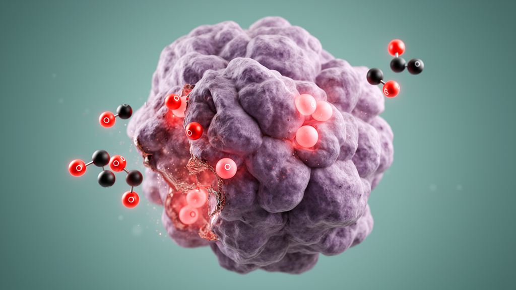
Researchers at the RIKEN Pioneering Research Institute in Japan have demonstrated a proof-of-concept technique that hijacks the excess carbon monoxide produced inside tumor cells and uses it to synthesize toxic anti-cancer compounds directly at the site of disease. The study, led by Katsunori Tanaka of the Biofunctional Synthetic Chemistry Laboratory and published in Nature Communications, describes a synthetically designed compound that enters cancer cells, exploits their internal carbon monoxide as a chemical feedstock, and triggers the production of a cytotoxic drug that then kills the very cells that made it. Because healthy cells do not carry the same surplus of carbon monoxide, the reaction—and the lethal compound it generates—is confined to the tumor.
Carbon monoxide is best known as the poisonous gas released by burning gasoline in car engines, but recent research has shown that many types of cancer cells also produce the molecule in elevated amounts. That makes carbon monoxide a potential biomarker of malignancy, provided it can be detected and exploited. Tanaka’s team set out to do exactly that, testing a system that delivers a metal catalyst into cells and pairs it with the intracellular carbon monoxide to assemble a bioactive compound. The chemistry at the heart of the approach relies on organometallic complexes called palladacycles, which can combine carbon monoxide into organic molecules to produce phenanthridinone. Several derivatives of phenanthridinone are cytotoxic and have already served as the basis for anti-cancer drugs. The obstacle was stability: a palladacycle placed naked inside a tumor cell is quickly deactivated by protective molecules within the cell, most notably the antioxidant glutathione, which destroys the palladium catalyst at the complex’s center before any useful synthesis can occur.
To shield the catalyst, the researchers drew on their lab’s specialty in in vivo metal-catalyzed reactions. They embedded the palladacycle inside albumin, an abundant protein that circulates naturally in the blood. The albumin–palladacycle complex can travel safely through the bloodstream, and once it enters a cell, the albumin shell protects the palladium from glutathione and other deactivating molecules. The team validated the system in stages. First, they screened a collection of palladacycles to identify the variant that produced the greatest yield of phenanthridinone. They then incubated the albumin–palladacycle complex for 48 hours with human liver, cervix, breast, and lung cancer cell lines, alongside a non-cancerous cell line as a control. After the incubation period, the desired reaction product was detected in the cancer cell lines but not in the normal cells, confirming both that the albumin wrapper protected the catalyst and that the reaction occurred selectively where excess carbon monoxide was present.
With targeting established, the researchers made adjustments to ensure the synthesized product detached from the complex and moved freely within the cell. They then modified the albumin–palladacycle complex so that the reaction generated a specific phenanthridinone derivative known to be highly toxic and already used in anti-cancer drugs. When tested in the liver cancer cell line, the team observed the drug being manufactured inside the cells, which subsequently suffered the toxic effects of the compound they had helped produce. One safety concern was carbon monoxide circulating in the blood, where accidental triggering of the catalyst during transport could have formed the toxic drug and destroyed blood cells before it ever reached a tumor. Testing showed, however, that the blood remained free of the drug—presumably because carbon monoxide in blood is bound to hemoglobin, rendering it unavailable for the palladium-catalyzed reaction. This built-in safeguard means the synthesis is effectively restricted to environments, like cancer cells, where carbon monoxide exists in a free, reactive form.
“This work represents an important advance in therapeutic in vivo synthetic chemistry,” Tanaka said. “In our therapeutic strategy, molecules naturally present or generated within the body—including disease-associated metabolites—are used as endogenous chemical feedstocks to synthesize therapeutic molecules directly at the site of disease.” In this concept, the active drug does not exist until the tumor’s own chemistry assembles it, an approach the researchers describe as selectively targeting tumors while avoiding harm to healthy cells. The findings remain at the proof-of-concept stage and were obtained in cultured cell lines rather than living organisms, so the approach has not yet been shown to work in animals or humans. The next step for the RIKEN team is to move from cell culture into animal models. If the technique can be extended beyond the laboratory, it could point toward treatments in which patients receive harmless precursor compounds that only become lethal drugs after being taken up by cancer cells.
Subject of Research: Cancer cells killed with their own carbon monoxide
Article Title: Cancer cells killed with their own carbon monoxide
Article References: Cancer cells killed with their own carbon monoxide. Original publication (Bibliographic details unavailable.)
Image Credits: AI Generated
DOI: Not provided
Keywords: Cancer, cells, killed, carbon, monoxide, scientific research
News Source: Nathaniel Bowman. (October 23, 2026). Cancer cells killed with their own carbon monoxide. Scienmag.