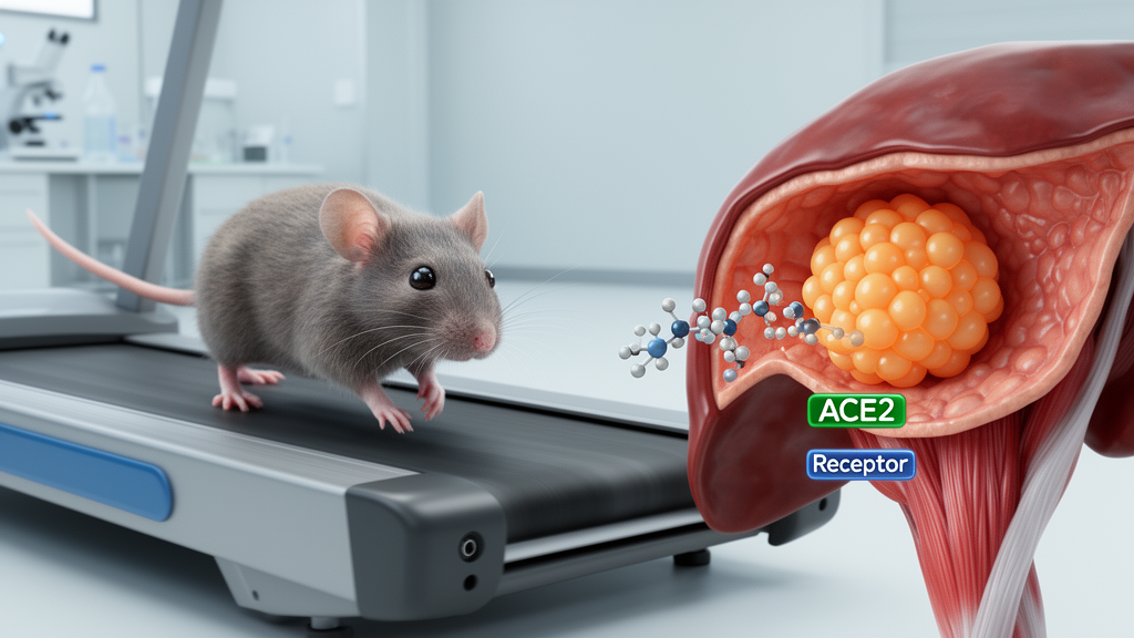
Aerobic exercise restored reduced levels of the mitochondrial peptide MOTS-c in the skeletal muscle, blood and liver of mice with metabolic dysfunction-associated steatotic liver disease (MASLD), according to a study in Scientific Reports. Giving MOTS-c directly also lowered liver fat, improved aspects of glucose metabolism and reduced inflammatory and fat-synthesis markers in the diabetic MASLD model. Experiments in liver cells and ACE2-knockout mice further indicated that the peptide acts through an AMPK–ACE2 pathway in the liver. The results identify a mechanistic link between exercise, muscle-derived signaling and liver metabolism, but they do not establish MOTS-c as an effective treatment for people with MASLD.
The study used a mouse model that combined type 2 diabetes with fatty liver. Male C57BL/6J mice received a high-fat diet for 16 weeks, and some animals also received injections of streptozotocin to induce diabetes; control animals received a standard diet. During the final four weeks, some diseased mice underwent daily aerobic training on a motorized platform, while others received MOTS-c at 7.5 mg/kg by intraperitoneal injection. The exercise regimen consisted of 60 minutes of running six days per week. Researchers measured MOTS-c concentrations and protein expression, glucose and insulin tolerance, circulating and liver lipids, liver histology, and molecular markers associated with fat synthesis, fatty-acid oxidation and inflammation. Separate experiments used male ACE2-knockout mice, and laboratory studies used HepG2 human liver cells exposed to palmitic acid as a metabolic stressor.
Mice with MASLD had lower concentrations of MOTS-c in serum, skeletal muscle and liver than normal controls. Aerobic training reversed these reductions, increasing skeletal-muscle expression and secretion and raising local MOTS-c levels in the liver. Administering MOTS-c directly also increased its expression in skeletal muscle, consistent with an autocrine effect, although exercise was more effective at elevating the peptide. The investigators proposed that aerobic activity may promote skeletal-muscle production and secretion of MOTS-c, which could then act on the liver. However, the experiment did not isolate the peptide’s contribution to all exercise effects: the training program altered several metabolic processes at once, and the investigators did not use a MOTS-c-specific knockout or antagonist.
After four weeks, both exercise and MOTS-c treatment lowered fasting blood glucose and reduced the area under the glucose- and insulin-tolerance curves in wild-type MASLD mice. Neither intervention significantly changed body weight. Exercise appeared more effective at restoring insulin sensitivity, whereas MOTS-c produced a marked improvement in glucose tolerance. The results therefore do not support treating the peptide and exercise as interchangeable. Their measured metabolic effects differed, and exercise increased the expression of ACOX1 and CPT-1, genes associated with fatty-acid beta-oxidation, whereas MOTS-c did not significantly increase these genes. Exercise was more effective at elevating MOTS-c than the injected peptide, which the authors attributed as a possible explanation rather than a directly tested mechanism.
Liver histology showed that both interventions reduced hepatic lipid accumulation. Serum and liver triglyceride concentrations fell, and serum low-density lipoprotein levels declined, while high-density lipoprotein levels did not differ significantly. At the molecular level, MOTS-c and aerobic exercise lowered expression of fatty acid synthase and acetyl-CoA carboxylase, which are involved in fatty-acid synthesis. They also reduced the inflammatory transcripts for interleukin-1 beta and tumor necrosis factor alpha. These changes accompanied less liver steatosis on hematoxylin-and-eosin and Oil Red O staining. Sirius Red staining and alpha-smooth-muscle actin staining did not indicate significant fibrosis in the experimental groups, meaning the study chiefly demonstrated effects on steatosis and metabolic or inflammatory markers rather than reversal of advanced liver scarring.
Hepatic ACE2 emerged as a molecular link between MOTS-c treatment and the improved liver phenotype. ACE2 expression was reduced in MASLD mice but was restored by either MOTS-c or exercise. The same general pattern appeared in HepG2 cells stressed with palmitic acid: ACE2 protein declined under metabolic stress and was partially restored by MOTS-c. ACE2 is a member of the angiotensin-converting enzyme family within the renin–angiotensin system, which the authors’ earlier research had linked to hepatic lipid balance. The present findings associate increased hepatic ACE2 with reduced steatosis and inflammation in the experimental model. The knockout experiments provided more direct evidence by testing what happened when ACE2 was absent.
That dependence was tested in mice lacking ACE2. In ACE2-knockout animals with MASLD, MOTS-c no longer significantly improved glucose or insulin tolerance, and its reductions in liver fat, triglycerides and expression of fat-synthesis and inflammatory genes were diminished or absent. By contrast, aerobic exercise still reduced hepatic steatosis and triglycerides in the knockout mice, altered fatty-acid synthesis and oxidation markers, and suppressed inflammatory gene expression. The divergence suggests that MOTS-c depends heavily on ACE2 for many of its measured effects in this model, while exercise engages additional mechanisms that do not require ACE2. It does not show that exercise acts exclusively through the peptide, and the study did not determine whether skeletal-muscle MOTS-c secretion or circulating peptide levels changed in the knockout animals.
The cell experiments placed phosphorylated AMPK, an energy-sensing kinase, upstream of the ACE2 response. Both MOTS-c and exercise increased phosphorylated AMPK in the livers of wild-type MASLD mice, while MOTS-c raised ACE2 and lowered fatty acid synthase protein. In palmitate-stressed HepG2 cells, MOTS-c similarly increased AMPK phosphorylation and ACE2 expression. Adding the AMPK inhibitor Compound C attenuated the MOTS-c-dependent increase in ACE2 and its suppression of fatty acid synthase and inflammatory genes. These findings support involvement of AMPK in the MOTS-c–ACE2 pathway. Pharmacological inhibition alone does not identify the precise molecular connection between AMPK activation and ACE2 regulation, however, and the authors did not establish whether the change involved ACE2 transcription, protein stability or another mechanism. They also proposed that transcriptional regulators such as SIRT1 or PPAR-gamma might participate, but that possibility was not tested.
Several design features limit how far the findings can be generalized. The investigators studied male mice and a human hepatoma cell line rather than patients with MASLD, and there were no human trial data. The MOTS-c intervention was administered by intraperitoneal injection, while exercise was delivered through a controlled platform-training regimen, so the routes and exposures were not directly equivalent. Small experimental groups, with several assays based on three to six animals, also constrain the precision of some estimates. The authors acknowledged the absence of a MOTS-c knockout mouse and a specific antagonist, which prevented direct loss-of-function testing of the peptide itself. They also noted potential safety concerns associated with systemic MOTS-c exposure, including possible effects on tumor progression and atherosclerosis. In addition, the model did not demonstrate significant fibrosis, leaving its relevance to advanced cirrhosis unresolved.
The broader significance is that the study offers a mechanistic explanation for one component of exercise’s metabolic effects: aerobic activity increased skeletal-muscle MOTS-c in diabetic MASLD mice, and the peptide was associated with activation of hepatic AMPK, restoration of ACE2 expression and reduced lipogenesis and inflammation. The intervention resembled exercise in improving steatosis and glucose regulation, but it did not reproduce every exercise effect. Its strongest metabolic contribution in the model was improved glucose tolerance, while exercise more clearly restored insulin sensitivity and promoted fatty-acid oxidation. Future translational studies would need to test whether the AMPK–ACE2 mechanism operates in people with MASLD, determine whether MOTS-c can be delivered safely and effectively, and establish which exercise protocols reliably increase endogenous production. Until such evidence is available, the results identify a promising pathway and experimental therapeutic target rather than a clinically established therapy.
Subject of Research: Experimental evidence linking exercise-induced skeletal-muscle MOTS-c to hepatic ACE2 and reduced MASLD features in mice and liver cells.
Article Title: Aerobic exercise induced skeletal muscle MOTS-c secretion: A novel therapy for MASLD via hepatic ACE2 regulation
Article References: Li, R., Li, F., Chen, Q., Gao, Y., Wu, Z., Deng, H., & Yuan, L. (2026). Aerobic exercise induced skeletal muscle MOTS-c secretion: A novel therapy for MASLD via hepatic ACE2 regulation. Scientific Reports, 16(1), Article 33291. https://doi.org/10.1038/s41598-026-60273-8
Image Credits: AI Generated
DOI: 10.1038/s41598-026-60273-8
Keywords: MOTS-c, MASLD, aerobic exercise, skeletal muscle, hepatic ACE2, AMPK, fatty liver, myokines, mouse model, liver metabolism, Aerobic, exercise
News Source: Denise Maddox. (October 22, 2026). Exercise-linked muscle peptide reduces fatty liver through ACE2 in mice. Scienmag.