Spinal cord injury remains one of medicine’s most devastating conditions, and yet one of its most familiar treatments—simply walking on a treadmill—may be doing far more than helping patients move again. A new study from researchers at the Federal University of Rio de Janeiro, published in the Journal of Cellular and Molecular Medicine, reveals that a carefully dosed treadmill rehabilitation protocol can fundamentally reshape the inflammatory environment inside an injured spinal cord, steering it away from destruction and toward repair. The findings, drawn from an unusually comprehensive set of behavioural, electrophysiological, molecular and imaging analyses, suggest that exercise is not merely a way to train surviving neural circuits but a biological intervention that actively quiets the secondary injury cascade.
The research team, led by Anne Caroline Rodrigues dos Santos Cavalcante and Suelen Adriani Marques, set out to answer a question that has lingered beneath years of encouraging rehabilitation data: when treadmill training improves locomotor recovery after spinal cord injury, what is actually happening inside the tissue? Previous work from the same group had shown that adjusting exercise volume according to the stage of injury maximises functional recovery, but the cellular and molecular mechanisms behind those behavioural gains remained largely opaque. To probe them, the researchers turned to a compressive injury model in fifty-two adult female mice, applying a 30-gram vascular clip to the spinal cord at the ninth thoracic vertebra for just ten seconds—a method that produces a reproducible, clinically relevant lesion while sparing the dura mater.
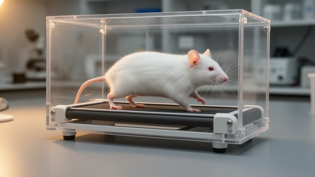
The rehabilitation protocol itself was deliberately conservative. Training began seven days after injury, during the subacute phase, and continued until fifty-six days post-injury. From day seven to day twenty-eight, animals completed two ten-minute treadmill sessions per training day, separated by a ten-minute rest interval; after day twenty-eight, this was reduced to a single ten-minute session. Sessions were held only three times per week, a frequency chosen to mirror what is actually feasible in clinical rehabilitation rather than to maximise exercise volume. The speeds ranged from six to twelve metres per minute, individually adjusted according to each animal’s locomotor performance and tolerance. This phase-adjusted design reflected the team’s earlier finding that exercise dosage must be tailored to the temporal evolution of the injury rather than simply intensified.
The behavioural results were striking. Untreated injured mice remained trapped in the earliest stage of locomotor recovery throughout the entire fifty-six-day follow-up, scoring just 1.48 on the Basso Mouse Scale by the end of the study. Treadmill-trained animals, by contrast, reached 4.02—a statistically significant difference that emerged from twenty-one days post-injury onward and persisted until the end of the experiment. On the Ladder Walking Test, which challenges animals to cross a horizontal ladder with irregularly spaced rungs, trained mice took significantly more total steps from thirty-five days post-injury and, more importantly, placed their paws significantly more accurately from forty-two days onward, indicating improved fine motor coordination rather than simply gross movement.
Sensory function also benefited. Using an electronic von Frey anesthesiometer, the researchers measured the pressure required to trigger a paw withdrawal reflex. Untreated injured animals showed markedly elevated thresholds—2.20 grams on the right paw and 2.01 on the left—reflecting profound sensory loss. Trained animals averaged 1.10 and 1.15 grams respectively, significantly closer to the uninjured baseline of roughly 0.61 grams. This partial preservation of sensory processing suggests that the rehabilitation protocol protected ascending pathways as well as descending motor tracts, a finding consistent with the broader tissue preservation documented throughout the study.
Electrophysiological recordings provided objective confirmation that neural conduction itself was preserved. At fifty-six days post-injury, the researchers exposed the cervical spinal cord and delivered supramaximal electrical stimuli while recording compound muscle action potentials from the gastrocnemius muscle. Untreated injured animals showed a dramatic reduction in signal amplitude—0.59 compared with 1.55 in uninjured controls—along with prolonged latency. Trained animals recorded amplitudes of 1.18, statistically indistinguishable from the uninjured group. Although such recordings do not directly demonstrate axonal regeneration, they provide compelling functional evidence that the descending motor pathways remained conductive in exercised animals, complementing the behavioural and morphological findings.
The molecular heart of the study lies in its analysis of the inflammatory microenvironment. Using ELISA assays on spinal cord tissue cultured for twenty-four hours, the team quantified six cytokines. Untreated injured animals exhibited a clear pro-inflammatory profile, with elevated levels of tumour necrosis factor-alpha, interleukin-1 beta and interleukin-6. Trained animals showed the opposite pattern: reduced pro-inflammatory cytokines alongside increased levels of the anti-inflammatory mediators interleukin-10, interleukin-13 and transforming growth factor-beta. This shift matters because sustained production of inflammatory cytokines after spinal cord injury drives microglial activation, immune cell infiltration, demyelination and neuronal apoptosis. During moderate exercise, skeletal muscle releases interleukin-6 as a myokine, which in turn promotes anti-inflammatory signalling and suppresses tumour necrosis factor-alpha production—a mechanism the authors cite as particularly relevant to their findings.
Immunofluorescence labelling revealed how this cytokine shift translated into changes in glial cell behaviour. The researchers used TMEM119 to mark the general microglial population, arginase-1 to identify anti-inflammatory M2-like microglia, and inducible nitric oxide synthase to flag pro-inflammatory M1-like cells. In trained animals, Pearson’s correlation between TMEM119 and arginase-1 reached 0.85, indicating that the majority of resident microglia had adopted an anti-inflammatory phenotype. In untreated injured animals, this correlation fell to 0.46, while the correlation between TMEM119 and the pro-inflammatory marker iNOS climbed to 0.836—compared with just 0.33 in trained mice. The team also documented reduced GFAP immunoreactivity in trained animals, suggesting attenuated reactive astrogliosis and a less restrictive glial scar, alongside significantly lower expression of cleaved caspase-3, a hallmark of apoptotic cell death.
Structural preservation followed the molecular changes. Transmission electron microscopy of semithin cross-sections at the lesion epicentre revealed that trained animals retained 1,658 myelinated nerve fibres on average, compared with just 200 in untreated injured animals. The g-ratio—the relationship between inner axonal diameter and total fibre diameter—shifted toward the optimal range of 0.6 to 0.7 in trained mice, indicating more efficient myelination. Micro-computed tomography confirmed that the lesion area was significantly smaller in trained animals (0.243 square millimetres versus 0.382), and scanning electron microscopy showed better organisation of both grey and white matter, with smaller cavitations at the injury site.
The benefits extended beyond the spinal cord itself. Trained animals preserved significantly more muscle mass in both the soleus and gastrocnemius, retained more neuromuscular junctions with their characteristic pretzel morphology, and maintained bladder wall thickness close to normal levels. Untreated injured animals developed dramatic bladder muscular hypertrophy—574.963 micrometres compared with 127.857 in controls—accompanied by inflammatory remodelling. The authors note that these autonomic complications are major contributors to urinary tract infections and repeated hospitalisation after human spinal cord injury. While the study used only female mice, a deliberate choice to reduce behavioural variability and avoid genital injury during recovery, the authors acknowledge that future work must include both sexes to confirm broader applicability. Taken together, the findings reframe treadmill rehabilitation not simply as locomotor training but as a coordinated biological therapy—one that appropriately dosed exercise can deploy to shift the injured spinal cord from a hostile inflammatory state toward one permissive for tissue repair and neural conduction.
Subject of Research: Neuroscience
Article Title: Treadmill Exercise Can Modulate the Inflammatory Microenvironment and Promote Regeneration After Compressive Spinal Cord Injury in Female Mice
Article References: Cavalcante, A. C. R. D. S., Laurindo, R. P., Heringer, L. D. S., Toledo, A. L. M. M., Souza, G. S., da Silva, G. F. Q., dos Santos, A. C. C., Branco, M. T. L. C., Martinez, A. M. B., & Marques, S. A. (2026). Treadmill Exercise Can Modulate the Inflammatory Microenvironment and Promote Regeneration After Compressive Spinal Cord Injury in Female Mice. Journal of Cellular and Molecular Medicine, 30(20), Article e71351. https://doi.org/10.1111/jcmm.71351
Image Credits: AI Generated
DOI: 10.1111/jcmm.71351
Keywords: spinal cord injury, treadmill exercise, neuroinflammation, microglia, rehabilitation, neuroregeneration, Treadmill, Exercise, Modulate, Inflammatory, Microenvironment, Promote
News Source: Cassandra Pierce. (October 17, 2026). Treadmill Training Rewires Inflammation to Heal Injured Spinal Cords in Mice. Scienmag.