Every second, somewhere in your body, a cell is dividing. In the lining of your intestine, this happens so frequently that the entire epithelial layer replaces itself every few days. For decades, biologists have treated cell division as a counting problem: one cell becomes two, the tissue grows, and the new arrivals somehow slot into place. A new study from the Stowers Institute for Medical Research, published online in Current Biology on September 30, 2026, reveals that this picture is radically incomplete. Cell division, it turns out, is not merely a way of adding cells. It is an active force that physically reshapes the tissue around it, choreographing the movements of neighbors in a precise and predictable sequence.
The research, led by former Stowers Postdoctoral Researcher Subu Ramanathan, Ph.D., now an Assistant Professor at the University of Nebraska Medical Center, under the direction of Stowers Investigator and Graduate School President Matt Gibson, Ph.D., focused on epithelial tissues. These are the tightly organized sheets of cells that cover body surfaces and line internal organs. Epithelia face a fundamental geometric dilemma every time they grow or renew themselves. A dividing cell must produce two daughter cells in a space that previously held only one, within a packed sheet where every cell is in contact with its neighbors. How the tissue accommodates this expansion without losing its architecture has been one of the enduring puzzles of tissue biology.
To watch the process unfold, the team turned to mouse intestinal organoids, miniature lab-grown models of gut tissue that recapitulate the structure of the intestinal lining. The intestine is one of the body’s most rapidly renewing tissues, making it an ideal system for studying how repeated rounds of cell division reshape tissue architecture over time. Crucially, organoids offered something that intact tissue rarely does: an unobstructed view. In most tissues, the basal surface of an epithelial cell, the side facing the underlying support layer, is hidden against neighboring cells or basement membrane. In intestinal organoids, that surface faces outward, giving the researchers an unusually clear window onto cell behavior that is normally difficult or impossible to observe directly.
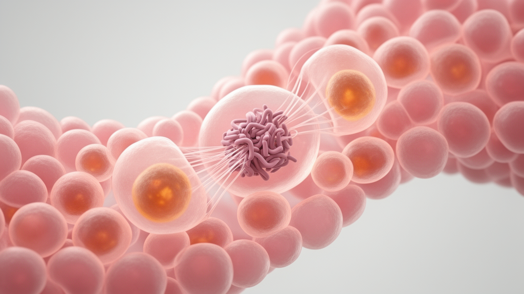
What Ramanathan saw when he first imaged the basal surface was striking. As cells divided, rosettes formed, flower-like clusters in which neighboring cells converged around the dividing cell, and neighboring cells visibly swapped positions with one another. Across 33 filmed cell divisions, the team documented the same overall sequence of rearrangements, and the live observations matched predictions from a computational model the researchers built alongside the imaging work. Together, the two approaches revealed that the dramatic shape changes cells undergo during mitosis, the process by which one cell splits into two, actively reorganize the surrounding tissue rather than passively accommodating it.
The sequence the researchers uncovered unfolds in distinct stages. As a cell prepares to divide, it first withdraws from the tissue’s basal surface, pulling away from the support layer. That withdrawal opens a temporary gap, and neighboring cells immediately close in, briefly forming the rosette structure. Then, as the two daughter cells reinsert themselves back onto the basal surface, they trigger a second round of rearrangement among the surrounding cells. The geometry of the returning daughters turns out to be the critical variable. Together, the two daughter cells were about 30 percent less circular than the original mother cell, producing a more elongated footprint that directed how neighboring cells reorganized, typically perpendicular to the line where the two daughter cells met.
The division process, as Ramanathan described it, creates a problem, a hole in a packed sheet, and then solves it through the geometry of the returning daughter cells. The shape change is not a side effect of mitosis. It does real work, actively sculpting the packing arrangement of the tissue. This insight emerged only after an initial, simpler hypothesis failed. The neighbor exchanges were not happening at the same time as the rosettes, which told the team their tidy story was incomplete. Ramanathan designed an experiment intended to destroy the hypothesis, and it did, revealing that withdrawal and reinsertion drive distinct parts of the remodeling process rather than a single coupled event.
The decisive experimental test came when researchers used a drug to arrest dividing cells before the daughter cells could return to the basal surface. The result was dramatic: neighbor exchange dropped by 93 percent, even though the rosettes still formed normally. This showed that the two phases of the process, withdrawal and reinsertion, are separable and serve different functions. Withdrawal alone creates the rosette, but it is the physical return of the elongated daughter cells that drives the actual swapping of neighbors. Blocking the second phase leaves the tissue with the gathering but without the reshuffling, demonstrating that mitotic geometry is the engine of the rearrangement.
The implications extend well beyond basic developmental biology. Up to 90 percent of cancer diagnoses in humans are carcinomas, cancers that arise from epithelial cell layers. Understanding how healthy epithelial tissues maintain their organization while adding cells may help researchers understand what goes wrong when abnormal growth disrupts that organization in disease. Gibson noted that the study adds to a growing body of work showing that epithelial proliferation is exquisitely choreographed. A dividing cell does not simply become two cells. Instead, the movements of mitosis create geometric changes that alter the organization of the tissue around it, meaning that cell division can act as a morphogenetic process, directly contributing to tissue form and not just tissue size.
The finding reframes how scientists think about proliferation at the tissue level. Cell division has conventionally been described as a mechanism for increasing cell number, with tissue shape maintained by separate remodeling mechanisms. The new work shows that these two functions are intertwined: the very act of division carries within it the instructions for local reorganization. Ramanathan put it plainly: we tend to think of cell division as simply making two cells from one, but this work shows that division also physically reshapes the tissue around it. Division is not just addition; it is a building process.
Many questions remain open. The researchers now want to understand how the physical properties of the surrounding tissue, including the basement membrane, work together with cell geometry during division, and whether the same rules hold across other growing and regenerating tissues. If the choreography observed in intestinal organoids proves to be a general principle, it could reshape how biologists think about development, wound healing, organ regeneration, and the early steps of tumor formation, all contexts in which tissues must grow while preserving, or losing, their precise architecture. For now, the study offers a striking reminder that one of biology’s most familiar events, the splitting of a single cell into two, is also one of its most elegant acts of structural engineering.
Subject of Research: How mitotic shape changes drive cell-contact rearrangements in proliferating epithelial tissue
Article Title: Stowers Institute scientists discover cell division does more than make new cells. It helps reshape tissues.
Article References: Stowers Institute scientists discover cell division does more than make new cells. It helps reshape tissues.. (n.d.). Original publication
Image Credits: AI Generated
DOI: Not provided
Keywords: cell division, epithelial tissue, tissue architecture, organoids, mitosis, Current Biology, Stowers Institute, morphogenesis, intestinal lining, cancer research, Stowers, Institute
News Source: Drew Townsend. (October 16, 2026). Cell Division Does More Than Make New Cells: It Helps Reshape Tissues. Scienmag.