Trypanosoma cruzi, the single-celled parasite behind Chagas disease, is one of the most genetically diverse human pathogens known, yet a new analysis suggests its core developmental biology is far more uniform than its tangled evolutionary history would imply. In a study published in Parasites & Vectors, researchers led by Víctor Hugo Caña-Bozada and Jaime A. Costales of the Pontificia Universidad Católica del Ecuador, together with colleagues at the Benemérita Universidad Autónoma de Puebla and the University of Glasgow, mined 86 publicly available RNA-seq libraries to chart the gene expression landscape of the parasite’s four major life cycle stages across multiple strains and genetic lineages. Their conclusion is striking: the transcriptional programs that define each developmental form are largely conserved, even across parasites belonging to different discrete typing units, the seven genetically distinct lineages into which the species is classified.
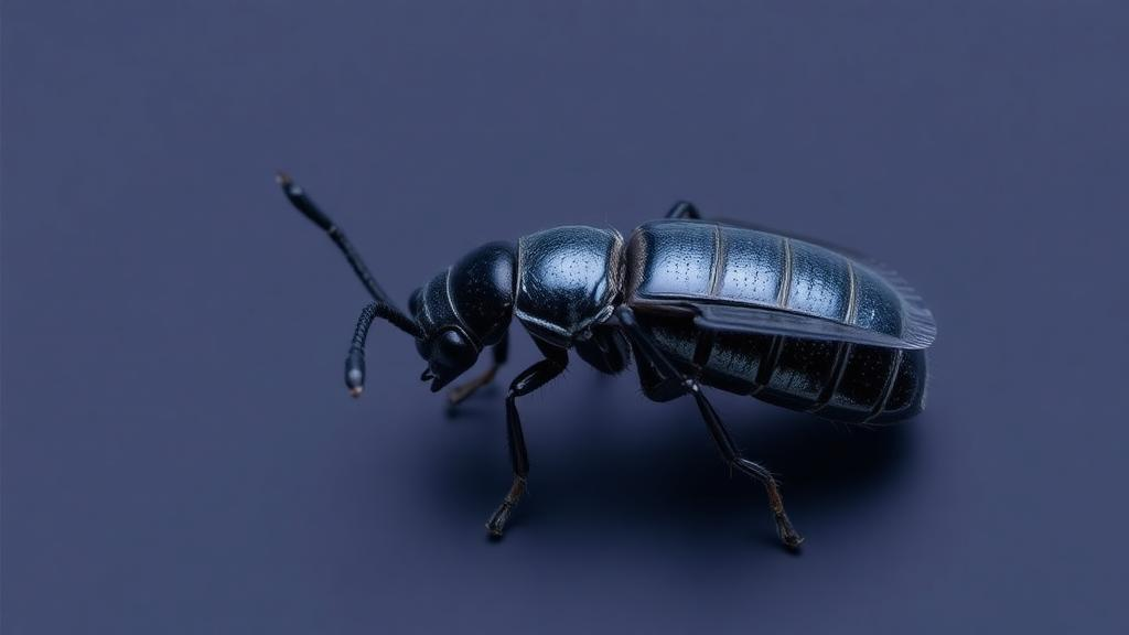
The parasite’s life cycle is a study in biological versatility. Inside triatomine bugs, the insect vectors sometimes called kissing bugs, T. cruzi exists as epimastigotes, replicating forms that multiply in the insect gut. As the parasite prepares to infect a mammalian host, it transforms into metacyclic trypomastigotes, the non-dividing forms deposited with insect feces that initiate infection. Once inside a mammal, the parasite invades cells and differentiates into amastigotes, the intracellular replicative stage, before converting back into tissue culture-derived trypomastigotes, the laboratory proxy for the blood form that circulates in the bloodstream and can be taken up by another feeding bug. Each of these forms must rewire its metabolism, surface chemistry, and cell biology to survive in radically different environments, and the new study set out to determine how much of that rewiring is shared across the species’ genetic spectrum.
Methodologically, the team faced a challenge familiar to anyone who has tried to combine public transcriptomic data: the libraries came from different laboratories, different strains, and different experimental protocols. The researchers filtered libraries derived from intracellular amastigotes to remove contaminating human reads, then processed all parasite data through a unified pipeline using Seq2Fun for cleaning, mapping, and quantification, which allowed them to work with orthologous groups rather than strain-specific gene models. Differential expression analysis was carried out with DESeq2, and functional interpretation drew on Gene Ontology and KEGG pathway enrichment using Gene Set Enrichment Analysis and over-representation analysis. The team also ran a parallel reference-genome mapping strategy with STAR and featureCounts for representative strains, including CL Brener, Colombia, and Y, and used co-expression network analysis and supervised classification to confirm that the biological signal was robust across bioinformatics approaches.
The results reveal a clear molecular identity for each stage. Epimastigotes, the replicative forms in the insect, show increased expression of genes tied to protein synthesis and energy metabolism, a signature of cells committed to growth and division. Notably, the same profile appears during the transitional phases as tissue culture-derived trypomastigotes convert into intracellular amastigotes, suggesting that the parasite reactivates a common replicative machinery whenever it needs to multiply, whether in a bug’s gut or inside a human cell. When the researchers compared tissue culture-derived trypomastigotes directly with epimastigotes, they identified a shared subset of 244 differentially expressed orthologous groups, a molecular common denominator pointing to shared mechanisms underlying these two distinct parasite forms.
The transitions in and out of the mammalian cell each carry their own conserved fingerprint. Differentiation into the amastigote form involves the overexpression of orthologous groups associated with metabolism, protein synthesis, and DNA repair, consistent with a stage that must both replicate and withstand the oxidative and nutritional stresses of the intracellular environment. The reverse transition, into tissue culture-derived trypomastigotes, activates functions linked to motility and signaling, and is marked by the overexpression of surface proteins, including mucins and trans-sialidases. These surface molecules are central to the parasite’s strategy for evading host immunity and invading new cells, and their coordinated upregulation at this stage underscores how the parasite arms itself for extracellular survival and onward transmission.
Yet the picture is not uniformly tidy. The metacyclic trypomastigote, the form responsible for transmitting infection from the insect vector to the mammalian host, stands out as the exception to the rule of conservation. In this stage, transcriptional signatures are strongly influenced by the parasite strain, a pattern the authors interpret as possible evidence of host adaptation. Because metacyclic trypomastigotes are the first forms to encounter a new mammalian host, strain-specific expression at this stage could reflect the fine-tuning of surface molecules and signaling pathways to particular transmission niches, an intriguing lead for understanding why different T. cruzi lineages are associated with different clinical outcomes and geographic distributions of Chagas disease.
The study also delivers a cautionary note for the laboratory community. Different in vitro methods for inducing metacyclogenesis, the artificial transformation of epimastigotes into metacyclic trypomastigotes, elicit divergent transcriptional programs in the parasite. The researchers compared metacyclic forms generated under different induction conditions and across strains, including the Colombia, Y, and MHOM lines, and found that the resulting expression profiles do not converge on a single consistent signature. Which experimental model best reflects the properties of metacyclic trypomastigotes in nature remains an open question, and the authors’ findings suggest that conclusions drawn from one induction protocol may not generalize to another, let alone to the parasite in the wild.
Beyond its immediate findings, the work provides something the field has lacked: a benchmark consensus resource. By integrating dozens of datasets from multiple strains and discrete typing units, including TcI, TcII, and TcVI, the authors have defined what they describe as the core standard landscape of the T. cruzi transcriptome. Future transcriptomic studies can now measure their own results against this consensus, distinguishing genuinely novel biology from artifacts of small sample sizes or idiosyncratic lab conditions. The analytical framework, which combines orthologous-group quantification with enrichment and network analyses, also offers a template for similar cross-strain comparisons in other genetically diverse pathogens.
The clinical stakes are considerable. Chagas disease affects millions of people across the Americas and is a leading cause of infectious heart disease, yet existing drug therapies are decades old, have limited efficacy in the chronic phase of infection, and carry significant side effects. Understanding which gene expression programs are conserved across the parasite’s genetic diversity helps identify the most stable targets for drug and vaccine development, since a therapeutic aimed at a universally expressed, essential pathway is less likely to fail against a divergent strain. Conversely, the strain-specific signature of the metacyclic stage hints at where the parasite’s adaptability resides, information that could inform surveillance and transmission-blocking strategies.
The study, funded by the UK Medical Research Council, is published open access, and its supplementary tables catalog the differentially expressed orthologous groups, enriched pathways, and co-expression modules in detail. As genomic datasets for T. cruzi continue to accumulate, this consensus transcriptomic map offers researchers a common reference point in a field where genetic diversity has often complicated comparison. For a parasite that shuttles between insect and mammal, replicates inside cells and in the bloodstream, and spans seven distinct lineages, the message of the new work is one of underlying order: the developmental playbook of T. cruzi, for the most part, is written once and shared by all.
Subject of Research: Stage-specific gene expression conservation across genetic lineages of the Chagas disease parasite Trypanosoma cruzi
Article Title: Stage-specific transcriptomic signatures in Trypanosoma cruzi are largely conserved across genetic diversity
Article References: Stage-specific transcriptomic signatures in Trypanosoma cruzi are largely conserved across genetic diversity. (n.d.). https://doi.org/10.1186/s13071-026-07689-3
Image Credits: AI Generated
DOI: 10.1186/s13071-026-07689-3
Keywords: Trypanosoma cruzi, Chagas disease, transcriptomics, RNA-seq, gene expression, parasite development, metacyclogenesis, discrete typing units, epimastigote, amastigote, trypomastigote, gene ontology
News Source: Juliet Wilcox. (October 14, 2026). Chagas Parasite Keeps Its Gene Expression Playbook Across Strains, Study Finds. Scienmag.