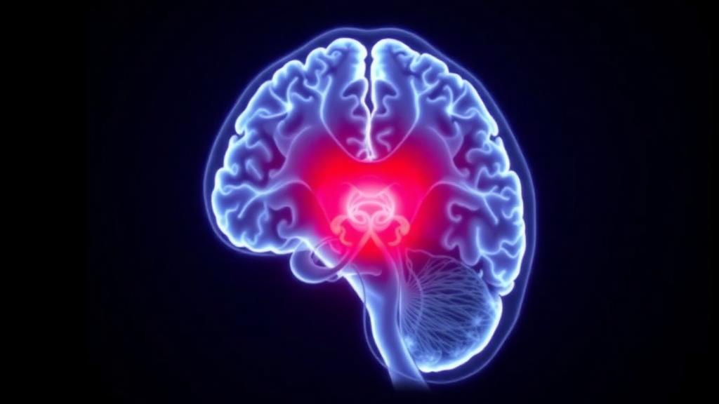
Radiation therapy saves lives in high-grade glioma, but it can also leave quiet scars on the healthy brain tissue it passes through. Now, a prospective imaging study from The University of Texas MD Anderson Cancer Center has captured those scars as they form, week by week, inside the hippocampus — the seahorse-shaped structure that anchors learning and memory. By scanning 45 patients every single week during their six-week course of partial-brain radiation, the researchers documented a progressive, dose-dependent shift in water diffusion within the hippocampus, offering one of the most detailed real-time portraits yet of how therapeutic radiation perturbs living human brain tissue.
The study, published in the Journal of Neuro-Oncology, enrolled adults with histologically confirmed high-grade glioma who were scheduled to receive the standard regimen of 60 Gy delivered in 30 fractions over six weeks, combined with the chemotherapy drug temozolomide. Nearly 89 percent of the cohort had glioblastoma, the most aggressive and common adult brain cancer. Before treatment began and then weekly throughout the course, each patient underwent diffusion magnetic resonance imaging on a 1.5-Tesla scanner, including high-resolution structural imaging and diffusion tensor imaging with 64 diffusion-encoding directions. A single experienced radiation oncologist manually contoured both hippocampi on every time point, avoiding the pitfalls of automated propagation, and extracted two quantitative metrics: the apparent diffusion coefficient, or ADC, and fractional anisotropy, or FA.
ADC measures how freely water molecules move through tissue. When cells are injured, membranes break down, the extracellular space expands, and water diffuses more easily — so ADC rises. FA, by contrast, quantifies how directionally organized the tissue microstructure is; in white matter tracts, demyelination and axonal damage cause FA to fall. Neither metric is specific to a single biological process, and the researchers were careful to frame them as nonsensitive but informative phenotypes of early tissue response rather than direct markers of irreversible injury. Still, the pattern they observed was striking in its consistency and its timing.
Hippocampal ADC climbed steadily from the very first week of treatment. The median relative increase was just 0.5 percent at Week 1, but it accelerated to 12 percent by Week 6, with an interquartile range of 6 to 18 percent. In a linear mixed-effects model, the time coefficient worked out to +2.2 percentage points per week, a highly significant trend. FA told a different temporal story: it remained statistically stable through the first three weeks and only dropped significantly during Weeks 4 through 6, from a baseline median of 0.181 to 0.165 at Week 6. Meanwhile, hippocampal volume barely budged — from a mean of 4.0 cubic centimeters at baseline to 3.97 at Week 6, a nonsignificant change. That dissociation is perhaps the study’s most intriguing clue: diffusion imaging appears to register radiation’s effects long before conventional structural measurements can detect any shrinkage.
Crucially, the magnitude of the diffusion change tracked the radiation dose each hippocampus received. After accounting for treatment week, every additional Gray of planned mean bilateral hippocampal dose was associated with an extra 0.81 percentage points of ADC change. Because the model included no dose-by-time interaction, this coefficient reflects a larger overall response with higher exposure rather than an accelerated weekly rate — but the dose association was robust. The team then sharpened the analysis by separating the hippocampus on the tumor’s side from the one opposite. Median planned dose was 15.5 Gy ipsilateral to the tumor versus only 6.7 Gy contralaterally, and the Week 6 ADC increase mirrored that asymmetry: 17 percent on the high-dose side versus 7 percent on the low-dose side. Yet dose-response relationships emerged independently in both hemispheres — 0.83 percentage points per Gy ipsilaterally and 0.36 contralaterally — suggesting the bilateral signal was not merely an artifact of averaging two very differently irradiated structures.
As a sanity check, the researchers examined a low-dose reference region, the left cerebellar tonsil, and found no association between dose and its ADC slope. They were careful to note this serves only as a limited internal reference rather than proof that the cerebellum is inherently radioresistant. Exploratory analyses also found no significant links between hippocampal ADC change and age, sex, steroid use, antiepileptic use, or broad tumor location, though the authors acknowledge these tests cannot exclude residual confounding.
The most provocative — and most preliminary — part of the study concerns memory. Twenty-four of the 45 patients completed both baseline and Week 6 cognitive testing with the Cogstate C3 battery, which includes tasks probing learning, face-name matching, and pattern separation. In exploratory, unadjusted comparisons, patients whose performance dropped by at least one standard deviation on the One Card Learning task showed a median ADC increase of 16.5 percent, compared with 10 percent in those who remained stable or improved. The composite memory score told a similar story: 18.5 percent versus 10 percent. FA change, by contrast, showed no significant association with any cognitive measure. The authors are explicit that these findings, drawn from a small subset with potential practice effects and no multiplicity correction, should not be read as validated thresholds or proof of lasting cognitive harm. But they sketch a testable framework — dose drives ADC response, which may relate to early memory change — that future studies can interrogate with larger samples, continuous cognitive outcomes, and longer follow-up.
The biological interpretation of a rising ADC during irradiation remains deliberately cautious. Increased water mobility could reflect expansion of the extracellular space from cellular and glial injury, disruption of membrane integrity, neuroinflammation, increased microvascular permeability, or simply shifts in tissue water content; in white matter, demyelination may contribute as well. The hippocampus is a particularly vulnerable target because its dentate gyrus hosts one of the brain’s few niches of adult neurogenesis, a process exquisitely sensitive to ionizing radiation. Prior work has linked hippocampal dose to long-term memory deficits: in adults treated with fractionated stereotactic radiation, and in children with medulloblastoma, where each additional Gray of mean hippocampal dose raised the odds of clinically meaningful associative-memory decline by roughly 23 to 26 percent. Other studies have traced dose-dependent diffusion changes in the corpus callosum and normal-appearing white matter during treatment, and dose-dependent hippocampal atrophy emerging about a year later. The new study extends this literature by resolving the hippocampal response itself, at weekly resolution, throughout a conventional treatment course — and by showing that early diffusion change and later structural atrophy may represent successive phases of the same continuum.
The limitations are real. The cohort was modest, dominated by glioblastoma, and the concurrent temozolomide means the observed changes cannot be attributed to radiation alone. Tumor proximity to the hippocampus, edema burden, seizure activity, and steroid exposure were not systematically quantified, and formal contour-reproducibility analyses were unavailable. The cognitive analyses, restricted to 24 completers who differed modestly from non-completers, remain exploratory in every sense. Yet the study’s core strength — repeated observation of normal-tissue response while radiation is actually being delivered — is hard to overstate. If early hippocampal ADC trajectories prove reproducible, particularly during Weeks 2 through 4, and if they predict later cognitive or volumetric outcomes beyond what dose and clinical factors already explain, they could become a quantitative imaging phenotype for adaptive treatment planning. Only then, the authors argue, should ADC-guided neuroprotective strategies be considered. For now, the study offers something rarer than a new therapy: a live, week-by-week view of the brain’s memory hub responding to treatment, captured before any damage becomes permanent on a conventional scan.
Subject of Research: Weekly diffusion MRI measurement of hippocampal microstructural changes and their dose dependence during partial-brain radiation therapy for high-grade glioma
Article Title: Weekly hippocampal diffusion changes during partial-brain radiation therapy for high-grade glioma: associations with dose and early memory change
Article References: Weekly hippocampal diffusion changes during partial-brain radiation therapy for high-grade glioma: associations with dose and early memory change. (n.d.). https://doi.org/10.1007/s11060-026-05822-x
Image Credits: AI Generated
DOI: 10.1007/s11060-026-05822-x
Keywords: high-grade glioma, glioblastoma, hippocampus, radiation therapy, diffusion MRI, apparent diffusion coefficient, fractional anisotropy, neurocognitive decline, memory, temozolomide, radiation dose, longitudinal imaging
News Source: Nathaniel Bowman. (October 13, 2026). Weekly MRI Reveals Radiation’s Early Fingerprint on the Brain’s Memory Hub. Scienmag.