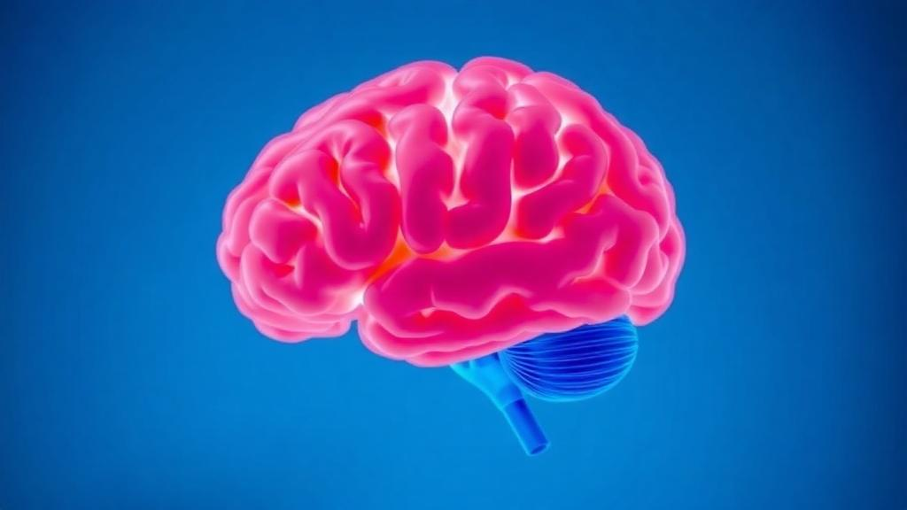
Older adults living with both type 2 diabetes and obesity may be showing the earliest invisible signs of brain stress decades before any diagnosis of dementia, according to a new pilot study published in the journal GeroScience. Using an advanced magnetic resonance imaging technique that can measure how hard the brain works to pull oxygen out of its own blood supply, researchers at the University of Texas Southwestern Medical Center found that participants with type 2 diabetes and obesity extracted oxygen more intensively across nearly the entire cerebrum than healthy, age-matched peers. The finding matters because type 2 diabetes is already known to raise dementia risk by roughly 55 to 65 percent in epidemiological studies, yet the precise mechanisms connecting disordered metabolism to failing cognition have remained stubbornly opaque. This study offers a fresh physiological clue: the brain’s oxygen extraction machinery appears to shift into a compensatory mode in people whose metabolism is under chronic strain.
The measurement at the heart of the study is called the oxygen extraction fraction, or OEF. In simple terms, OEF is the proportion of oxygen carried by the blood that is actually captured by tissue to keep cells functioning. In healthy brains, this value is remarkably stable across gray and white matter and varies little from person to person, which makes deviations potentially meaningful. When cerebral blood flow drops, the brain typically compensates by extracting a larger fraction of the oxygen that does arrive, a relationship governed by basic physiology and Fick’s principle, which links oxygen consumption to the product of blood flow and extraction. A rising OEF can therefore be read as a warning light on the dashboard: the plumbing may be struggling even if total energy use, measured as the cerebral metabolic rate of oxygen, has not yet declined.
What makes this study technically notable is that the team measured OEF without contrast agents or radiation, using quantitative susceptibility mapping, an MRI method that converts tiny magnetic field disturbances caused by deoxygenated hemoglobin into quantitative maps of venous oxygenation. The researchers acquired multiecho gradient-recalled echo images on a 3 Tesla Siemens Prisma scanner, unwrapped the phase data with an open-source algorithm, removed background field contributions, and inverted the local field maps into susceptibility values using a morphology-enabled dipole inversion approach. Cerebrospinal fluid in the ventricles served as a zero reference, and a deep learning model based on an attention-gated 3D U-Net segmented cortical veins automatically, achieving a Dice coefficient of about 0.79 in held-out testing. Partial volume effects were corrected with an orientation-aware cylindrical fitting method, and subject-specific hematocrit values from blood draws were plugged into the biophysical equation linking susceptibility to oxygen saturation.
The study enrolled 32 participants aged 55 to 85, split into 14 older adults with type 2 diabetes and obesity and 18 age- and sex-matched controls. The diabetes group had a body mass index between 30 and 45 kilograms per square meter and hemoglobin A1c values between 6.0 and 10 percent, while controls had normal-range BMI and HbA1c below 5.7 percent. Anyone with prior stroke, major neurological or psychiatric illness, heart disease, or uncontrolled hypertension was excluded, and the two groups were statistically indistinguishable in age, sex, and education. On average, the participants with diabetes had been diagnosed about 5.7 years earlier and were, by clinical standards, fairly well controlled, with a mean HbA1c of 6.7 percent. Their insulin resistance, however, measured by the HOMA-IR index, was markedly elevated at 5.6 compared with 2.2 in controls, well above a commonly cited metabolic syndrome threshold.
The headline result was striking in its consistency. Whole-brain OEF averaged 29.5 percent in the diabetes and obesity group versus 27.3 percent in controls, a relative increase of about 8 percent that survived statistical correction for multiple comparisons. Every lobar region examined, including the frontal, temporal, parietal, occipital, and insular cortices, showed the same elevation. The largest gap appeared in the occipital lobe, where OEF reached 37.0 percent in the metabolic group against 33.3 percent in controls, an 11 percent relative difference. Crucially, cerebral blood flow measured with arterial spin labeling was mostly similar between groups, and the cerebral metabolic rate of oxygen, calculated by combining flow and extraction, did not differ in any region. That combination, higher extraction with preserved consumption, is exactly the signature expected when the brain is compensating for vascular stress rather than suffering outright metabolic failure.
The cognitive testing added a sobering dimension. Although all scores fell within clinically normal ranges, the diabetes and obesity group performed significantly worse on the Repeatable Battery for the Assessment of Neuropsychological Status, with lower scores in list learning, coding, attention, and the global cognition index. They also scored markedly lower on the Color-Word Switching condition of the Delis-Kaplan Executive Function System, a demanding test of inhibitory control and mental flexibility, where the difference was the largest observed. Depression scores on the Geriatric Depression Scale were equivalent between groups, arguing against mood as a confound. The pattern, spanning attention, processing speed, and executive function rather than a single domain, overlaps with what clinicians see in early vascular cognitive impairment, the spectrum of thinking difficulties caused by compromised brain blood supply.
Even more intriguing were the correlations between brain physiology and behavior. Across all participants, higher regional OEF was consistently associated with poorer cognitive performance, with the strongest relationships appearing in the temporal lobe, where OEF correlated at minus 0.62 with executive switching performance, and minus 0.41 with attention. The direction was uniformly negative across every region tested. On the metabolic side, whole-brain OEF rose with blood leptin levels, a hormone produced by fat tissue that is elevated in obesity and linked to adipose dysfunction and neuroinflammation, and fell with higher HDL cholesterol, the lipoprotein widely regarded as cardioprotective. HbA1c and fasting insulin showed positive trends that approached significance. These associations are exploratory and were not corrected for multiple comparisons, so the authors are careful to frame them as hypothesis-generating rather than definitive.
The interpretation favored by the researchers is that elevated OEF represents an early compensatory response to metabolic and vascular stress, a kind of cerebral overdrive that precedes measurable declines in resting brain metabolism. This fits with prior work showing that OEF is elevated in people with heavy vascular risk burdens, particularly those with cognitive impairment, and with evidence that the occipital lobe is a common site of reduced perfusion in type 2 diabetes. It also echoes findings in other conditions, from asymptomatic carotid artery disease to sickle cell anemia and obstructive sleep apnea, where altered oxygen extraction tracks with cognitive difficulty. If confirmed, OEF could serve as a sensitive, quantitative, and entirely noninvasive marker of cerebrovascular vulnerability, potentially distinguishing vascular contributions to dementia risk from purely neurodegenerative ones, something that visually rated markers like white matter hyperintensities cannot do with the same objectivity.
The caveats are real and the authors state them plainly. This was a small, single-center, cross-sectional pilot study, so it cannot establish causation, cannot disentangle the independent effects of diabetes from those of obesity, and cannot rule out that differences in medication use, including higher rates of antihypertensive treatment in the metabolic group, contributed to the imaging findings. The exploratory statistical analyses carry an elevated risk of false positives. What the study does establish is a plausible and testable physiological link: older adults with type 2 diabetes and obesity show diffusely elevated cerebral oxygen extraction, and that elevation tracks with both adverse metabolic markers and weaker attention and executive performance. The next step, the researchers say, is larger longitudinal cohorts that follow OEF, blood flow, and cognition over time to determine whether this compensatory signature predicts who will decline, and whether interventions that improve metabolic health can normalize the brain’s oxygen economy before damage becomes permanent.
Subject of Research: Cerebral oxygen extraction fraction measured by quantitative susceptibility mapping in older adults with type 2 diabetes and obesity and its associations with cognition and metabolic markers
Article Title: Regional oxygen extraction fraction is higher in older adults with type 2 diabetes and obesity: associations with cognition and metabolic markers
Article References: Elsharydah, M. N., Al-Khoury, Y., Ragunatha Rao, S., Schaffert, J., Saha, A., Kaufman, J., Rezaei, A., Leary, J., Jin, A., Almandoz, J. P., Gauthier, C. J., & Thomas, B. P. (2026). Regional oxygen extraction fraction is higher in older adults with type 2 diabetes and obesity: associations with cognition and metabolic markers. GeroScience. https://doi.org/10.1007/s11357-026-02590-4
Image Credits: AI Generated
DOI: 10.1007/s11357-026-02590-4
Keywords: type 2 diabetes, obesity, oxygen extraction fraction, quantitative susceptibility mapping, cerebral blood flow, cognitive impairment, dementia risk, GeroScience, MRI biomarker, insulin resistance, leptin, vascular cognitive impairment
News Source: Daisy Hatcher. (October 13, 2026). Diabetes and Obesity May Force the Aging Brain to Work Harder for Oxygen. Scienmag.