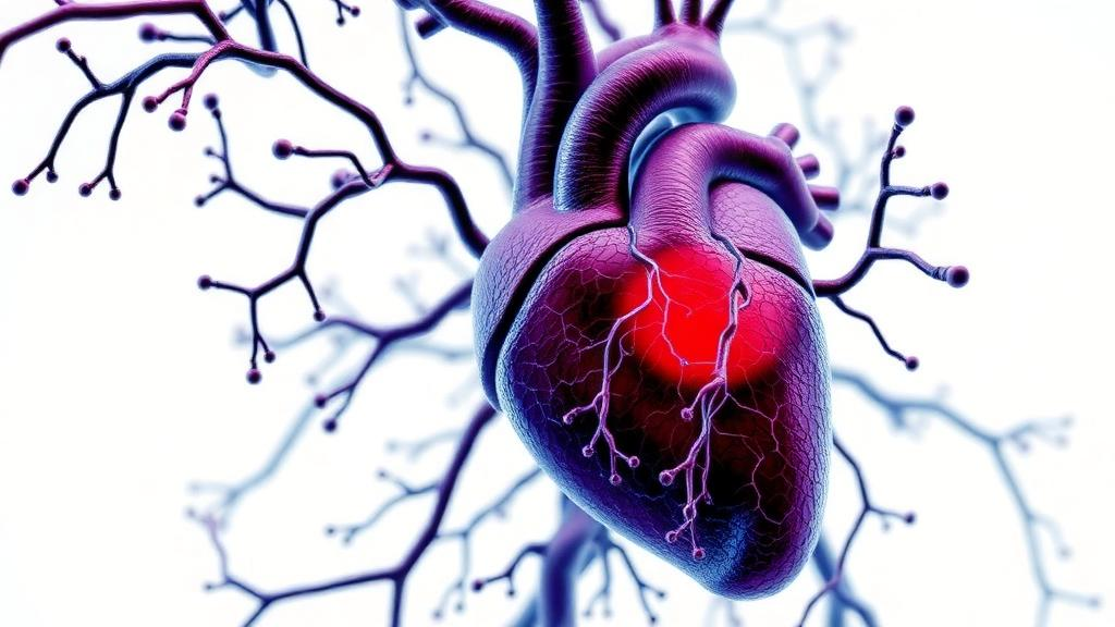
The heart has long been treated by immunologists as a bystander, an organ caught in the crossfire of generic inflammatory processes rather than a specific target of the adaptive immune system. A new review published in Nature Reviews Cardiology by Gustavo Campos Ramos of the University Hospital Würzburg argues that this view is overdue for revision. Drawing together decades of work in experimental models and a rapidly growing body of human data, the review makes the case that T cells directed against cardiac proteins participate in a striking range of conditions, from acute myocarditis and myocardial infarction to chronic heart failure, and that harnessing this antigen-specific crosstalk could open the door to precisely targeted therapies. The obstacle, the author contends, is definitional: only a small number of cardiac antigens have been conclusively identified, and the field still lacks a rigorous immunological definition of what makes an immune response heart-specific in the first place.
Much of modern cardioimmunology has been built on the premise that inflammation matters, but that its specificity does not. That premise has produced real clinical wins. Large trials of canakinumab, an interleukin-1beta antibody, demonstrated that blunting innate inflammation reduces cardiovascular events in patients with atherosclerotic disease, and low-dose colchicine has entered routine secondary prevention. More recently, randomized phase 2 trials of low-dose interleukin-2, intended to expand regulatory T cells, have tested whether boosting immune tolerance can calm vascular and myocardial inflammation after acute coronary syndromes. Imaging probes that track CCR2-positive monocytes into the injured heart have given clinicians a window onto the inflammatory infiltrate. Yet all of these approaches share a fundamental limitation: they target broad inflammatory programs rather than the antigen-specific lymphocytes that may be orchestrating, sustaining or sometimes protecting the myocardium.
The evidence that antigen-specific T cell responses to the heart exist is not new. In the late 1980s, researchers showed that immunizing genetically predisposed mice with cardiac myosin, the molecular motor of the heartbeat, induced autoimmune myocarditis, and subsequent work established that the disease was T cell mediated. The myocarditis-inducing epitope of cardiac myosin heavy chain alpha was mapped and shown to bind stably to major histocompatibility complex class II molecules on antigen-presenting cells. Decades later, the clinical relevance of these findings crystallized around a dramatic iatrogenic disease: myocarditis triggered by immune checkpoint inhibitors, the cancer therapies that unleash T cells against tumors. Independent teams showed that T cells specific for alpha-myosin and other cardiac myosin epitopes expand in patients with checkpoint-inhibitor myocarditis, providing the clearest demonstration yet that a heart antigen can drive fulminant human cardiac autoimmunity.
Why would the immune system be primed against its own heart? Part of the answer lies in central tolerance. The thymus deletes T cells that react strongly with self-proteins it displays, but the gene encoding alpha-myosin heavy chain, the dominant cardiac contractile protein, is expressed poorly or not at all in thymic medullary epithelial cells. This gap in thymic tolerance, demonstrated in both mice and humans, leaves a reservoir of T cells capable of recognizing the heart’s most abundant autoantigen. Peripheral mechanisms normally keep these cells in check, but injury can break the truce. When cardiomyocytes die, as in a heart attack or viral infection, cardiac proteins are released, taken up by dendritic cells and presented to T cells in a pro-inflammatory context, a process known as cross-priming. Molecular mimicry adds another route: microbial peptides that resemble cardiac epitopes, whether from group A streptococci in rheumatic heart disease or from the gut microbiota in experimental inflammatory cardiomyopathy, can activate T cells that then attack the heart.
The catalogue of validated cardiac antigens remains short but instructive. Cardiac myosin heavy chain alpha dominates, with defined class II-restricted epitopes and, more recently, identified class I epitopes for CD8 T cells. Cardiac troponin I is a second major player; unlike troponin T, it induces severe autoimmune inflammation in the myocardium when used to immunize mice, and autoantibodies against it correlate with outcomes after infarction. The beta-1 adrenergic receptor, long known as a target of functional autoantibodies in dilated cardiomyopathy, also contains epitopes that elicit T cell responses of varying pathogenicity. Additional candidates include the adenine nucleotide translocator, SERCA2a, the calcium pump of the sarcoplasmic reticulum, and branched-chain alpha-ketoacid dehydrogenase kinase. Beyond classical self-antigens, modified self can be antigenic: isolevuglandin-modified cardiac proteins drive CD4 T cell activation and dysfunction in the heart, and citrullination of myofilament proteins may reveal cryptic epitopes, a mechanism familiar from rheumatoid arthritis.
What these antigens share, and what the review proposes as a working framework, is a combination of abundance, cardiac enrichment and presentability. The heart is overwhelmingly composed of cardiomyocytes packed with contractile machinery, so its proteome is dominated by sarcomeric proteins that are scarce elsewhere. Proteomic atlases of the human heart, from quantitative maps of ventricular and atrial regions to single-cell and spatial transcriptomics of cardiac niches, now provide the raw material for predicting which proteins are sufficiently heart-enriched and sufficiently abundant to serve as targets. The critical filter is immunological: a candidate antigen must actually be processed and presented on MHC molecules. Strikingly, myosin-class II complexes have been detected in normal myocardium even before any injury, suggesting that the heart constitutively displays its own dominant antigen and that disease depends on context rather than mere exposure.
Not all heart-directed T cell responses are destructive, and this nuance may prove therapeutically decisive. Myocardial infarction has been shown to trigger cardioprotective, antigen-specific T helper responses, and a unique population of cardiac regulatory T cells potentiates protection after ischemic injury. In atherosclerosis, T cells reactive against apolipoprotein B begin as protective regulatory cells and can lose that identity as disease progresses, converting from guardians to drivers of plaque inflammation. Regulatory T cells that recognize troponin or myosin epitopes modulate healing after infarction, and the myocardial milieu itself favors local differentiation of regulatory T cells. The same antigen, in other words, can support repair or pathology depending on the functional polarization of the T cells that recognize it, a duality that any antigen-specific intervention must navigate carefully.
Turning this biology into medicine will require tools to see the relevant T cells, and such tools are finally emerging. MHC multimers loaded with defined cardiac peptides allow direct staining and isolation of antigen-specific T cells from blood and tissue. Single-cell T cell receptor sequencing, applied to paired atherosclerotic plaques and blood, has revealed expanded effector clones with autoimmune features, and comparable approaches have mapped the immune response in checkpoint-inhibitor myocarditis across heart, blood and tumor. Curated databases of pathology-associated TCR sequences, machine-learning methods for inferring TCR specificity, and CRISPR-based orthotopic TCR replacement for validating candidate receptors together form a toolkit that the review positions as the foundation for the next decade of mechanistic work. Transgenic mice carrying myosin-specific T cell receptors, first generated more than a decade ago, continue to illuminate how CD4 and CD8 subsets contribute to myocarditis and its progression to dilated cardiomyopathy.
The therapeutic horizon is equally provocative. Engineered T cells targeting fibroblast activation protein have been shown in preclinical studies to blunt cardiac fibrosis, including chimeric antigen receptor T cells produced in vivo via mRNA delivery, hinting at a future where the antigen-specific arm of immunity is redirected rather than suppressed. Conversely, costimulation blockade with abatacept has been deployed as rescue therapy for severe checkpoint-inhibitor myocarditis, a rational application of the two-signal model of T cell activation to a fulminant, antigen-driven disease. Preventive vaccine-style approaches that tolerize against cardiac autoantigens have shown promise in experimental heart failure. None of these strategies can be designed rationally, the review concludes, without a complete inventory of cardiac antigens and the T cell repertoires that recognize them. By fusing cardiac proteomics with immunopeptidomics, the systematic analysis of peptides displayed on MHC molecules, and with T cell receptor tracking from model organisms to patients, the field aims to convert heart-specific immunity from a scattered collection of observations into a defined, druggable axis of cardiovascular medicine.
Subject of Research: Antigen-specific T cell responses to cardiac proteins in myocarditis, myocardial infarction and heart failure
Article Title: Heart-specific T cell responses: from antigens to disease mechanisms
Article References: Campos Ramos, G. (2026). Heart-specific T cell responses: from antigens to disease mechanisms. Nature Reviews Cardiology. https://doi.org/10.1038/s41569-026-01356-x
Image Credits: AI Generated
DOI: 10.1038/s41569-026-01356-x
Keywords: T cells, cardiac antigens, myocarditis, myocardial infarction, heart failure, autoimmunity, MHC, immunopeptidomics, cardioimmunology, regulatory T cells, immune checkpoint inhibitors, T cell receptor
News Source: Kristina Jarvis. (October 12, 2026). T Cells That Recognize the Heart: Mapping the Antigens Behind Cardiac Disease. Scienmag.