A team of chemical engineers in Shanghai has developed a precisely controlled way to manufacture tiny porous spheres made of polycaprolactone, a biodegradable polymer already familiar to cosmetic medicine, and shown that the porous versions coax human skin fibroblasts into producing more collagen than their smooth-surfaced counterparts. The work, published in the Archives of Dermatological Research, offers both a manufacturing recipe and a biological rationale for why the surface architecture of injectable filler particles might matter as much as the material they are made from.
Dermal fillers have evolved well beyond the simple gels that dominated the market for decades. A newer generation of so-called biostimulatory fillers does not merely occupy space under the skin; it is designed to provoke the patient’s own cells to rebuild the collagen scaffold that aging progressively dismantles. Polycaprolactone, often abbreviated PCL, has emerged as one of the leading materials in this class. It is slow to degrade, mechanically robust, and has a track record in approved collagen-stimulating products. Yet the microscopic details of how PCL particles interact with fibroblasts, the workhorse cells of the dermis, remain only partly understood, and manufacturers have largely relied on batch emulsion methods that produce particles with wide, poorly controlled size distributions.
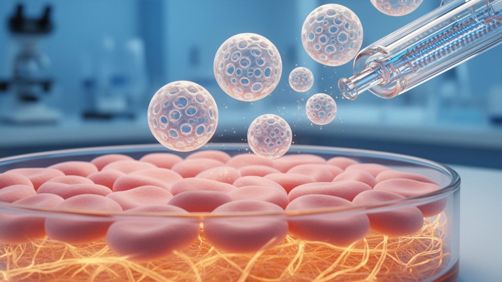
The research group, led by Jintao Zhang and Lian Cen at East China University of Science and Technology, turned to droplet-based microfluidics to close that gap. In a microfluidic device, liquids are pushed through channels narrower than a human hair, and the interplay of flow rates, channel geometry and surface tension determines exactly when and how droplets pinch off. Because each droplet becomes one particle, the technique yields spheres of remarkably uniform diameter, a property known as monodispersity that conventional stirring-based emulsification cannot match. In this study, the researchers tuned the ratio of flow rates in the device to produce PCL microspheres of around 49 micrometers in diameter, a size range considered suitable for injection through fine needles while remaining large enough to persist in tissue.
The crucial innovation was the introduction of porosity. The team used tridecane, a simple hydrocarbon, as a porogen, a sacrificial agent that creates voids in the solidifying polymer. As the PCL droplets hardened, the tridecane was expelled in a way that left behind an interconnected porous architecture. The resulting porous microspheres, designated PM in the study, achieved a porosity of 78.19 percent, meaning more than three quarters of their apparent volume consisted of open space. For comparison, the researchers also prepared compact microspheres, designated SM, with smooth, dense surfaces and the same average size, allowing them to isolate the effect of surface texture from that of particle size or chemistry.
With both particle types in hand, the team subjected them to a battery of cell culture experiments using human skin fibroblasts, the cells responsible for synthesizing the collagen matrix that gives young skin its strength and elasticity. Both porous and compact microspheres proved highly biocompatible: fibroblasts grew on and around them, proliferated robustly, and showed no signs of the toxicity that can plague synthetic biomaterials. But the porous particles consistently outperformed their compact counterparts at the same concentration, stimulating greater fibroblast proliferation and, critically, greater secretion of both type I and type III collagen, the two fibrillar collagens most relevant to dermal structure and repair.
The dose mattered as well. The researchers systematically varied the concentration of microspheres in culture and identified 0.5 milligrams per milliliter as the optimal concentration for promoting collagen secretion. Below that level, the stimulatory effect was weaker; above it, no additional benefit was observed. This kind of dose-response mapping is exactly the information that translational developers need when formulating injectable products, since the concentration of particles in a filler suspension directly influences both its rheology and its biological activity after injection.
One of the more nuanced findings concerned inflammation. Interleukin-6, a signaling molecule often cast as a marker of harmful inflammation, was progressively upregulated in fibroblasts treated with either type of microsphere, with the porous particles producing a slightly stronger effect than the compact ones. The authors interpret this within the framework of regenerative signaling rather than pathology: transient, moderate IL-6 expression is part of the normal wound-healing and remodeling program, and many biostimulatory materials work precisely by engaging these pathways. The study thus adds a piece to the puzzle of how microsphere fillers trigger neocollagenesis, the formation of new collagen, without settling the deeper question of how to balance stimulatory inflammation against chronic irritation.
The implications reach beyond cosmetic dermatology. Porous micro- and nanoparticles are under intense investigation across regenerative medicine as cell carriers, drug depots and tissue-engineering scaffolds, and topographical cues, the physical texture of a biomaterial surface, are increasingly recognized as powerful regulators of cell behavior, sometimes rivaling biochemical signals. By demonstrating that a single manufacturing variable, surface porosity, measurably changes collagen output from human fibroblasts, the study strengthens the case that filler design should be treated as a materials-science problem as much as a formulation problem. It also aligns with parallel work on porous microspheres of poly-L-lactic acid, another biostimulatory polymer, which have been reported to act faster and produce fewer nodules than dense formulations.
The microfluidic platform itself may prove to be the study’s most exportable contribution. Because particle size in a droplet microfluidic device is governed by tunable flow rate ratios, the same hardware could, in principle, produce filler particles matched to different clinical indications: smaller spheres for superficial rhytids, larger ones for deep volume restoration. Monodispersity also promises more predictable degradation kinetics and more consistent biological response from batch to batch, addressing a longstanding criticism of emulsion-produced microsphere products. The authors describe their work as providing both a superior functional filler candidate and a detailed manufacturing technology, and the combination is what gives the paper its practical weight.
Considerable distance remains between cell culture dishes and clinic. The experiments reported here were conducted in vitro with a single cell type, and the immune environment of living skin, with its macrophages, vasculature and mechanical loading, will shape the fate of injected particles in ways no fibroblast monoculture can fully predict. Long-term studies of degradation, migration and nodule formation will be essential before porous PCL microspheres can be proposed for human use. Still, the study delivers something the field has lacked: a clean, controlled demonstration that porosity itself, engineered through microfluidics, amplifies the collagen-stimulating capacity of a proven filler polymer. As demand grows for fillers that regenerate tissue rather than simply fill it, manufacturing precision of this kind is likely to become the standard against which next-generation products are judged.
Subject of Research: Microfluidic fabrication of porous polycaprolactone microspheres as collagen-stimulating dermal fillers
Article Title: Microfluidic preparation of porous polycaprolactone microspheres as dermal fillers with enhanced collagen secretion
Article References: Zhang, J., Li, Y., Ye, J., & Cen, L. (2026). Microfluidic preparation of porous polycaprolactone microspheres as dermal fillers with enhanced collagen secretion. Archives of Dermatological Research, 318(1), Article 436. https://doi.org/10.1007/s00403-026-04944-6
Image Credits: AI Generated
DOI: 10.1007/s00403-026-04944-6
Keywords: polycaprolactone, microfluidics, porous microspheres, dermal fillers, collagen synthesis, fibroblasts, skin aging, biostimulatory fillers, tissue engineering, interleukin-6, biomaterials, regenerative medicine
News Source: Eric Holt. (October 9, 2026). Porous Polymer Microspheres Made by Microfluidics Boost Collagen in Skin Cells. Scienmag.