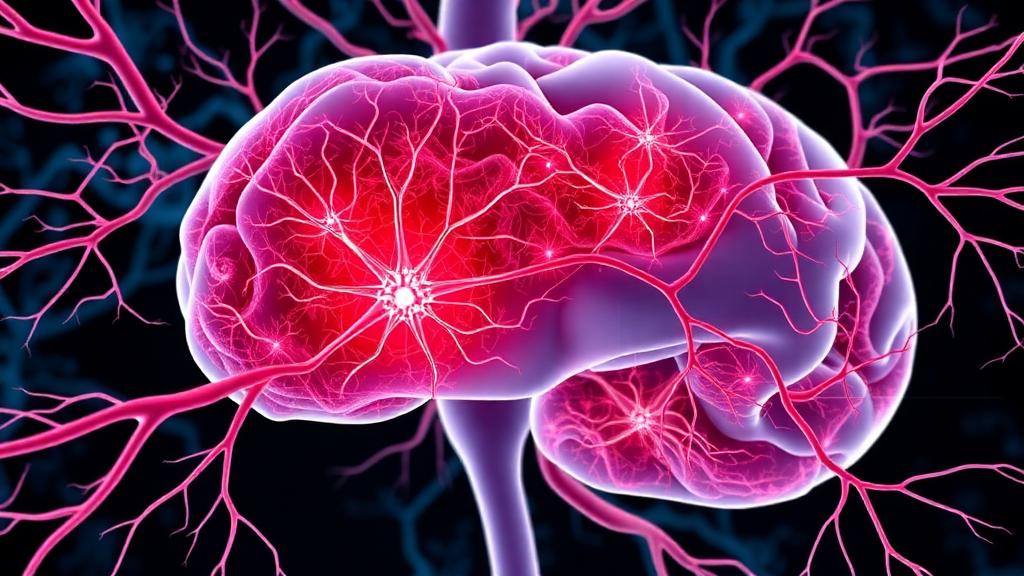
Hepatocellular carcinoma, the most common form of primary liver cancer, remains one of the world’s deadliest malignancies, and clinicians have long struggled to explain why some tumors behave far more aggressively than their size or stage would suggest. A new review published in the Journal of Translational Medicine argues that a long-overlooked culprit may be hiding in plain sight: the nerves that thread through and around the tumor. The paper, authored by Meijing Yi, Jingting Zhang, Zhuo Liu, Wei Peng, Wenhui Gao, Renyi Yang and Puhua Zeng of Hunan University of Chinese Medicine and affiliated institutions, synthesizes the scattered clinical evidence for perineural invasion in liver cancer and proposes a detailed mechanistic framework for how tumor cells and nerve fibers may fuel one another’s growth. The work arrives at a moment when the emerging field of cancer neuroscience is rapidly redrawing the boundary between the nervous system and malignancy.
Perineural invasion, often abbreviated PNI, describes the process by which cancer cells surround, track along and eventually infiltrate nerve trunks. It is a well-recognized hallmark of aggressive behavior in pancreatic, prostate and head and neck cancers, where it correlates with recurrence, metastasis, disease-specific death and debilitating neuropathic pain. In hepatocellular carcinoma, however, the picture has been muddled. The review’s authors note that the prevalence and prognostic relevance of PNI in primary liver cancer remain inconsistently defined, with limited and heterogeneous clinical data making it difficult to judge whether nerve invasion is a common driver of the disease or a rare epiphenomenon. This uncertainty, they argue, is itself a problem worth solving, because tumor–nerve crosstalk is now known to modulate tumor growth, immune remodeling, dissemination and resistance to therapy.
The clinical stakes are considerable. Hepatocellular carcinoma is a major cause of cancer mortality worldwide, and most patients are diagnosed at advanced stages when curative options have narrowed. If neural invasion proves to be a genuine feature of the hepatic tumor microenvironment, it could help explain patterns of local recurrence and spread that current staging systems fail to capture. It could also open an entirely new axis of treatment. Drugs that interrupt neurotransmitter signaling or block neurotrophic pathways are already in clinical use for other conditions, and repurposing them against the tumor–nerve interface is an attractive translational prospect. But the authors are careful to temper expectations: without improved clinical and experimental rigor, causality and actionable targets cannot be established.
To bring order to a fragmented literature, the review organizes the potential drivers of perineural invasion in hepatocellular carcinoma into five domains: neurotrophic factors, axon guidance molecules, neurotransmitter signaling, cell adhesion molecules and the broader tumor microenvironment. The first domain centers on the chemical attractants that nerves themselves produce and respond to. Nerve growth factor, brain-derived neurotrophic factor and glial cell-derived neurotrophic factor, together with the GDNF family ligands, can be secreted by tumor cells and stromal cells alike, drawing axons into the tumor mass through receptors such as TrkA and TrkB, the p75 neurotrophin receptor and the GDNF family receptor alpha complex paired with the RET tyrosine kinase. Once engaged, these receptors activate downstream signaling cascades involving phosphoinositide 3-kinase, protein kinase B, the MAPK pathway and the transcription factors STAT3 and NF-κB, promoting tumor cell survival, proliferation and epithelial-mesenchymal transition.
Axon guidance molecules form the second domain, and their role in cancer is among the most intriguing findings of modern tumor biology. Semaphorins, slits and ephrins were originally characterized as traffic controllers that steer growing axons during development, repelling or attracting nerve fibers with exquisite precision. The review highlights how hepatocellular carcinoma cells may co-opt these same ligand-receptor systems, including semaphorin signaling through neuropilin-1, SLIT ligands engaging Roundabout receptors, and ephrin signaling through EphA2, to migrate along neural highways. In effect, the tumor borrows the embryo’s navigation toolkit, following chemotactic gradients that normally guide nervous system wiring. Matrix metalloproteinases secreted along the way degrade the extracellular matrix, clearing a physical path for both tumor cells and advancing axons and creating a mutually reinforcing corridor of invasion.
The third domain, neurotransmitter signaling, connects the tumor to the body’s systemic physiology. Adrenergic signaling through norepinephrine and epinephrine acting on alpha- and beta-adrenergic receptors can elevate cyclic AMP, activate protein kinase A and the cAMP-responsive element-binding protein, and thereby influence tumor proliferation and stress adaptation. Cholinergic signaling is equally consequential: acetylcholine acting through muscarinic receptors such as M1R and through the alpha-7 nicotinic acetylcholine receptor modulates inflammatory circuits within the tumor. The review also points to metabolic crosstalk, including signaling through the bile acid receptor TGR5, as a hepatically specific wrinkle that may distinguish liver cancer’s neural niche from those of other tumors. Because stress, circadian disruption and systemic inflammation all reshape neurotransmitter flux, this domain links a patient’s whole-body state to the molecular behavior of the tumor.
The fourth and fifth domains emphasize physical contact and ecological context. Cell adhesion molecules provide the mechanical grip that allows tumor cells to cling to the perineurium, the protective sheath surrounding nerve fibers, and to crawl along it with relative impunity, shielded from many conventional therapies. The tumor microenvironment completes the picture. Cancer-associated fibroblasts, tumor-associated macrophages and regulatory T cells secrete chemokines such as CCL2 and CXCL family ligands, along with cytokines like interleukins, transforming growth factor-beta and vascular endothelial growth factor, that recruit both nerves and immunosuppressive cells into the same spatial niche. Hypoxia, driven by hypoxia-inducible factor-1 alpha, further reshapes this landscape. The authors describe these neuro-immune-stromal circuits as creating permissive perineural niches, microenvironments in which nerve fibers, cancer stem cells and suppressive immune cells sustain one another in a self-reinforcing loop.
What distinguishes this review from a simple catalog of molecules is its attention to experimental infrastructure. The authors outline a series of platforms specifically designed to test their hypotheses: co-culture systems that pair hepatocellular carcinoma cells with neurons and Schwann cells, the glial cells that wrap peripheral nerves; three-dimensional organoids that recapitulate the architecture of the hepatic tumor niche; dorsal root ganglia-based assays that quantify axonal attraction; and microfluidic nerve-on-chip models that allow researchers to observe tumor-nerve interactions in real time under controlled gradients of chemokines and neurotransmitters. Such tools, the review argues, are essential because standard two-dimensional cultures cannot capture the geometry, mechanics and multicellular complexity of perineural invasion. Dedicated hepatocellular carcinoma-focused PNI models, the authors stress, simply do not yet exist at the level of sophistication the field requires.
The review’s most sobering conclusion concerns the state of the clinical evidence itself. Standardized pathological assessment of perineural invasion in liver resection specimens is lacking, and reporting practices vary so widely across studies that meta-analytic conclusions are fragile. The authors call for harmonized diagnostic criteria, dedicated HCC-focused models and reproducible experimental designs before the field can move from hypothesis to therapy. Their caution is well placed: cancer neuroscience has generated abundant correlative findings across tumor types, but converting nerve-tumor crosstalk into validated drug targets demands exactly the kind of mechanistic and clinical rigor this review prescribes. Still, the direction of travel is clear. If the neural niche proves as influential in liver cancer as the molecular evidence suggests, the sympathetic and parasympathetic nervous systems may join angiogenesis and immune evasion as central characters in the story of hepatocellular carcinoma, and the beta-blockers, neurotrophin antagonists and axon guidance inhibitors already sitting on pharmacy shelves may find an unexpected second act in oncology.
Subject of Research: Perineural invasion mechanisms and clinical significance in hepatocellular carcinoma
Article Title: Perineural invasion in hepatocellular carcinoma: clinical evidence and mechanistic hypotheses
Article References: Yi, M., Zhang, J., Liu, Z., Peng, W., Gao, W., Yang, R., & Zeng, P. (2026). Perineural invasion in hepatocellular carcinoma: clinical evidence and mechanistic hypotheses. Journal of Translational Medicine. https://doi.org/10.1186/s12967-026-08910-8
Image Credits: AI Generated
DOI: 10.1186/s12967-026-08910-8
Keywords: hepatocellular carcinoma, perineural invasion, cancer neuroscience, tumor microenvironment, neurotrophic factors, axon guidance molecules, neurotransmitter signaling, tumor-nerve crosstalk, nerve-on-chip models, organoids, epithelial-mesenchymal transition, Journal of Translational Medicine
News Source: Nathaniel Bowman. (October 7, 2026). Nerves and Tumors Talk: New Review Maps How Liver Cancer Invades Neural Highways. Scienmag.