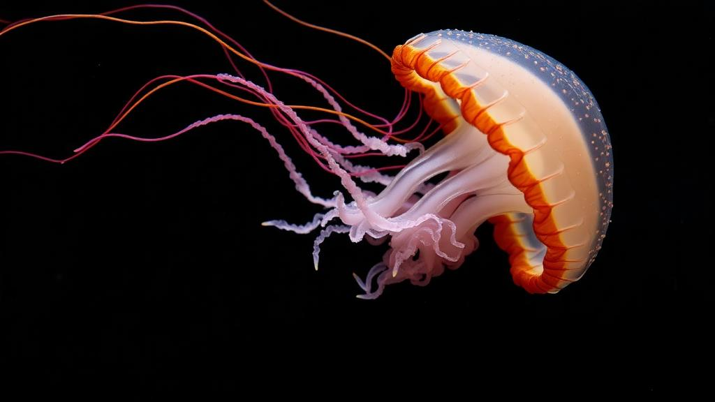
Regeneration has long been celebrated as one of biology’s most astonishing abilities, from axolotls regrowing entire limbs to flatworms rebuilding themselves from fragments. Yet a new study of a small hydrozoan jellyfish suggests that even animals endowed with remarkable regenerative powers cannot escape the grip of aging. Researchers investigating the medusa stage of the cnidarian Cladonema pacificum have shown that growing older progressively dismantles the cellular machinery required for tissue maintenance and repair, offering some of the clearest evidence yet that aging constrains regeneration even in early-branching animals that sit far from the vertebrate lineage on the tree of life.
Cladonema pacificum has recently emerged as a tractable laboratory model for studying regeneration in medusae, the free-swimming, sexually reproducing stage of the cnidarian life cycle. Unlike the sessile polyp Hydra, which can display negligible senescence under laboratory conditions, medusae follow a finite and reproducible lifespan, making them an ideal system in which to ask how aging reshapes tissue homeostasis within a single life-history context. The jellyfish retains robust regenerative capacity during early adulthood, so the researchers could track precisely when and how that capacity begins to fail. Their findings, published in Aging Cell, reveal a coordinated breakdown across multiple cellular systems rather than a single point of failure.
The team began by monitoring medusae under standardized culture conditions, taking longitudinal measurements of the three major organs: the umbrella that powers swimming, the manubrium that handles feeding and gametogenesis, and the tentacles used for prey capture and defense. As the animals aged, all three structures deteriorated. Umbrellas shrank significantly, manubria regressed relative to body size, and tentacles became markedly shorter. The researchers classified animals up to 40 days old as young, those between 41 and 60 days as middle-aged, and those 61 days and beyond as aged, a scheme grounded in the consistent, measurable changes they observed across dozens of individuals.
Histological analysis deepened the picture of decline. In young medusae, the manubrium displays a well-organized bilayered structure with oocytes nestled in its interior; in aged animals, the outer ectodermal layer thinned dramatically and oocyte numbers appeared reduced. Egg production, tracked longitudinally in the same individuals, peaked during the young phase and then fell steadily. The tentacles told a similar story: the photoreceptor organs called ocelli, neatly circular in youth, became irregular and disrupted with age, while the spherical clusters of stinging nematocytes at the tentacle tips grew less prominent. Critically, the accumulation of phosphorylated histone H2AX, a widely used molecular marker of DNA damage, rose in the tentacle bulbs as the animals aged, linking the visible morphological decay to cellular-level damage.
Because tentacles are the best-characterized regenerative tissue in Cladonema, the researchers focused their cellular analysis there. Hydrozoan tentacles harbor multipotent interstitial stem cells, or i-cells, concentrated near the tentacle base, which act as resident homeostatic stem cells supplying both progenitors and differentiated cells. Using fluorescence in situ hybridization and immunostaining, the team found that mature nematocytes, identified by the poly-γ-glutamate they synthesize, declined significantly in aged tentacles. Their precursors, the nematoblasts marked by the genes Mcol1 and Nanos2, were also reduced, and an independent, marker-independent histological scoring of the nematoblast-rich region confirmed the shrinkage of this lineage. Neuronal cell bodies near the tentacle base, labeled with anti-FMRFamide staining, likewise dropped in number.
The stem cells themselves showed clear signs of functional decline. Quantification based on β-catenin staining and nuclear morphology revealed a modest reduction in the proportion of i-cell-like cells in aged tentacle bulbs, alongside an increased share of epithelial cells, and FISH for the stem cell marker Piwi showed a marked drop in Piwi-positive cells. Proliferation collapsed across the board: EdU incorporation assays revealed far fewer S-phase cells in aged bulbs, and phospho-histone H3 staining showed fewer mitotic figures. Notably, when the researchers combined EdU labeling with Piwi FISH, they found that DNA synthesis declined both in Piwi-positive stem-like cells and in the broader Piwi-negative population, indicating that the proliferative slowdown was not confined to the stem cell compartment but swept through the entire tissue.
Did this cellular erosion translate into regenerative failure? To find out, the team amputated tentacles from medusae aged 30 to 70 days and monitored regrowth. Young tentacles elongated steadily, formed branches, and rebuilt their nematocyte clusters, but regeneration grew progressively weaker with age, with the decline becoming especially pronounced between 50 and 60 days. Functional recovery fared even worse. Seven days after amputation, every regenerated tentacle in young medusae captured and ingested prey, whereas most aged tentacles failed to capture prey at all and none completed ingestion. Even the earliest step of regeneration, wound closure driven by remodeling of the actin cytoskeleton, was substantially delayed: while most young tentacles achieved complete epithelial closure within two days, few aged tentacles managed it even by three days.
The mechanistic culprit appears to be the blastema, the localized mass of proliferative, undifferentiated cells that assembles at the wound site and orchestrates regrowth. Previous work established that Cladonema blastema formation depends on repair-specific proliferating cells, a lineage largely distinct from the resident homeostatic stem cells, and that blocking proliferation anywhere in the system impairs regeneration. In the aged medusae, EdU incorporation at wound sites was severely reduced, mitotic cells were scarce, and FISH revealed far fewer Piwi- and Nanos1-positive cells accumulating at injuries. Morphology-based classification confirmed a loss of i-cell-like cells near aged wounds. Intriguingly, a substantial population of morphology-defined stem-like cells remained at the injury sites even in old animals, but these cells had largely lost the molecular markers and proliferative activity that define a functional blastema, suggesting that aging impairs the ability of existing cells to enter the regenerative state rather than simply depleting them.
The authors interpret blastema failure not as a primary cause of regenerative decline but as an integrated outcome of broader aging defects: reduced baseline proliferation, weakened injury responses, and deteriorating stem cell support all precede or accompany the blastema’s collapse. They also note that changes in the stem cell niche, the local tissue environment that provides instructive cues, may contribute alongside intrinsic changes in the stem cells themselves. Because regeneration-specific cell lineages are distinct from homeostatic ones, aging may selectively compromise the activation or coordination of repair programs, perhaps through alterations in cellular state or the surrounding tissue architecture.
The comparative implications are striking. In the African turquoise killifish, aging disrupts progenitor activation and early regenerative events in brain and fins; in axolotls, limb regeneration slows with age rather than disappearing outright. Cladonema now extends this pattern to a cnidarian, an animal lacking dedicated mobile immune cells and classical inflammatory responses, implying that aging targets core components of regenerative systems, stem and progenitor cell dynamics and their coordination with surrounding tissues, across the breadth of metazoan evolution. The study also joins a growing body of work showing that strong regenerative capacity does not guarantee escape from senescence: sexual Hydra oligactis deteriorates after induced reproduction, and sexual planarians show clear aging despite their legendary plasticity. Cladonema, with its short life cycle, easy laboratory culture, and experimentally accessible tentacles, now offers researchers a powerful new platform for dissecting how the biology of aging collides with the biology of repair, and why, in the end, regeneration alone cannot hold back the tide of time.
Subject of Research: Age-related decline in tissue homeostasis and blastema-mediated tentacle regeneration in the cnidarian medusa Cladonema pacificum
Article Title: Aging Disrupts Tissue Homeostasis and Constrains Blastema‐Mediated Regeneration in the Cladonema Medusa
Article References: Kanehisa, R., Nakatani, H., Takatori, S., Tomita, T., Miura, M., & Nakajima, Y.-I. (2026). Aging Disrupts Tissue Homeostasis and Constrains Blastema‐Mediated Regeneration in the Cladonema Medusa. Aging Cell, 25(10), Article e70727. https://doi.org/10.1111/acel.70727
Image Credits: AI Generated
DOI: 10.1111/acel.70727
Keywords: Cladonema pacificum, aging, regeneration, blastema, stem cells, cnidarians, medusa, tissue homeostasis, nematocytes, DNA damage, proliferation, hydrozoa
News Source: Beatrice Stafford. (October 6, 2026). Aging Undermines Regeneration Even in the Remarkable Jellyfish Cladonema. Scienmag.