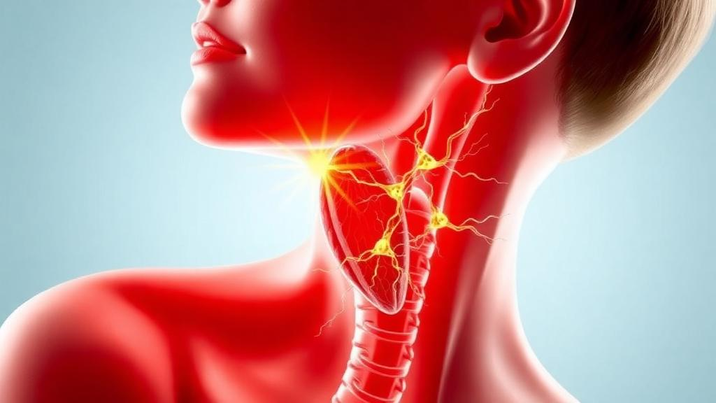
For decades, clinicians assumed that the dramatic weight loss seen in patients with head and neck squamous cell carcinoma was largely a mechanical problem: tumors and their treatments make swallowing painful and difficult, so patients simply eat less. A new review published in Supportive Care in Cancer argues that this explanation, while partially true, misses the deeper story. Drawing together evidence from tumor biology, immunology, and imaging, researchers led by Vincenzo Zurlo and Francesco Perri of the Istituto Nazionale Tumori IRCCS Fondazione G. Pascale in Naples propose that cancer-related malnutrition in head and neck cancer is best understood as a tumor-driven immune-metabolic syndrome, in which the malignancy itself actively dismantles the patient’s body from the inside out.
The biological centerpiece of this framework is metabolic reprogramming, one of the updated Hallmarks of Cancer. Head and neck squamous cell carcinoma cells rely heavily on aerobic glycolysis, the so-called Warburg effect, burning through enormous quantities of glucose and converting it to lactate even in the presence of oxygen. This inefficient ATP generation might seem like a design flaw, but it allows rapid proliferation and creates a continuous drain on the body’s nutrient supply. The lactate released by tumors does not simply disappear; it enters the Cori cycle, forcing the liver to spend energy converting it back into glucose through gluconeogenesis. The result is a futile substrate loop that raises resting energy expenditure and contributes to the hypermetabolic state long observed in cancer patients.
The review emphasizes that the tumor behaves, in the authors’ framing, as a metabolic sink. Glucose, lipids, and amino acids are redirected from skeletal muscle, liver, and adipose tissue toward the growing malignancy. Cytokine-driven activation of proteolytic pathways in muscle triggers the breakdown of contractile proteins and the release of glutamine and other amino acids that can be funneled into tumor metabolism. Meanwhile, tumor-derived factors such as parathyroid hormone-related protein promote lipolysis and the browning of white adipose tissue, converting energy stored in fat into heat rather than usable fuel. These processes, the authors stress, do not directly nourish the tumor; instead, they force the host to mobilize substrates in a losing attempt to maintain homeostasis, producing thermogenic inefficiency and accelerating energy wasting.
Chronic systemic inflammation amplifies this catabolic storm. Elevated circulating levels of interleukin-6, tumor necrosis factor-alpha, and interleukin-1 beta are commonly found in head and neck cancer patients and correlate with weight loss, sarcopenia, and poorer clinical outcomes. Interleukin-6 has been strongly linked to loss of body mass and adipose tissue depletion, while tumor necrosis factor-alpha enhances ubiquitin-proteasome-mediated protein degradation and impairs glucose transport in peripheral tissues. Within skeletal muscle, activation of the myostatin/SMAD signaling pathway suppresses protein synthesis and accelerates atrophy. Together, these mechanisms create a self-perpetuating loop in which inflammation sustains catabolism, tissue wasting further fuels inflammatory signaling, and the patient’s metabolic reserve erodes progressively, often before any anticancer therapy has begun.
This model explains a clinical observation that has long puzzled oncologists: many patients lose weight and muscle mass early in the disease course, even when their oral intake appears adequate, and caloric supplementation alone frequently fails to prevent muscle loss or cachexia. The internationally accepted GLIM criteria for diagnosing malnutrition already recognize inflammation as a key etiologic driver, requiring at least one phenotypic criterion such as weight loss or reduced muscle mass alongside an etiologic criterion such as reduced intake or disease-related inflammation. The new review goes further, arguing that in head and neck cancer the inflammatory and metabolic components dominate, and that nutritional impairment should be viewed as a dynamic, tumor-driven process rather than a simple caloric deficit.
Translating this biology into measurable clinical tools is where the review makes its most distinctive contribution. Sarcopenia, the progressive loss of skeletal muscle, is one of the most clinically relevant manifestations of cancer-related metabolic derangement. Because patients with head and neck cancer rarely undergo routine abdominal imaging, cross-sectional muscle assessment at the level of the third cervical vertebra has emerged as a validated surrogate for whole-body muscle mass. The resulting C3 skeletal muscle index can identify muscle depletion that body mass index misses entirely, and it carries prognostic weight: sarcopenia has been consistently associated with poorer overall survival, increased treatment-related toxicity, and reduced quality of life, making it a robust biomarker rather than a mere byproduct of treatment intolerance.
On the metabolic side, functional imaging offers a window into the tumor’s glycolytic appetite. Total lesion glycolysis, derived from 18F-FDG PET scans, integrates tumor volume with metabolic activity to reflect the overall glucose consumption of the malignancy. High values have been associated with aggressive tumor biology, greater inflammatory burden, and adverse oncologic outcomes. Crucially, recent prospective data in locally advanced head and neck squamous cell carcinoma showed that higher tumor metabolic activity measured by total lesion glycolysis was significantly associated with reduced skeletal muscle mass at the third cervical vertebra, elevated inflammatory markers, and impaired nutritional status at the start of treatment. This provides clinical validation of the biological model: tumor-driven hypermetabolism and inflammation translate into measurable deterioration of body composition and nutritional reserve.
The authors are careful to acknowledge the limitations of the current evidence base. The studies linking PET-derived metabolic parameters with nutritional impairment remain limited in number and observational in design, and external validation in larger prospective cohorts is still needed. Methodological heterogeneity in PET acquisition protocols, lesion segmentation strategies, and cutoff values complicates comparisons across studies, and standardized imaging protocols and universally accepted thresholds for C3-based muscle assessment have not yet been fully established. Metabolic imaging biomarkers should therefore be considered complementary tools alongside conventional nutritional and body composition assessment rather than stand-alone diagnostic markers. In practice, the review advocates a multimodal approach combining clinical nutritional evaluation, C3-derived muscle assessment when available, inexpensive and widely accessible inflammatory markers such as C-reactive protein and the neutrophil-to-lymphocyte ratio, and metabolic imaging parameters in patients already undergoing PET imaging.
The therapeutic implications are perhaps the most consequential part of the framework. If malnutrition is an immune-metabolic syndrome, then early nutritional intervention should aim not merely to increase calories but to modulate systemic inflammation and preserve functional body composition. Current European Society for Clinical Nutrition and Metabolism guidelines support early nutritional intervention as a cornerstone of multidisciplinary care in head and neck oncology, and intervention appears most effective during the pre-cachectic or early cachectic phases, when metabolic alterations remain potentially reversible. Immunonutrition enriched with arginine, omega-3 fatty acids, and nucleotides has shown promising effects, including improvement of the CD4/CD8 T-cell balance and accelerated postoperative recovery, but the evidence remains heterogeneous, and the authors caution that adequately powered randomized trials are needed before routine use beyond current guideline indications can be recommended.
Patients with locally advanced disease, high tumor metabolic burden, marked systemic inflammation, and early muscle depletion are especially vulnerable, as are those with advanced age, HPV-negative tumors, extensive smoking histories, dysphagia, or treatment with concurrent chemoradiotherapy. For these individuals, the review argues, nutritional management should prioritize preservation of lean body mass over weight maintenance alone, with adequate protein and energy provision embedded in integrated supportive care that includes pain control and oncologic treatment planning. Nutritional support alone is unlikely to fully reverse the underlying immune-metabolic alterations, but early recognition of high-risk features could shift the paradigm from reactive nutritional rescue to proactive metabolic management. By uniting tumor metabolism, systemic inflammation, and imaging-based body composition into a single conceptual framework, the Naples team offers oncologists a way to identify vulnerable patients before cachexia takes hold, and a biological rationale for treating the tumor’s metabolic sabotage, not just the patient’s empty plate, as the true target of nutritional care in head and neck cancer.
Subject of Research: Tumor-driven immune-metabolic mechanisms of cancer-related malnutrition in head and neck squamous cell carcinoma
Article Title: Beyond reduced intake: tumor metabolism and immune dysregulation drive cancer-related malnutrition in head and neck squamous cell carcinoma (HNSCC)
Article References: Zurlo, V., Piccirillo, A., Marciano, M. L., Pontone, M., Rampetta, F. R., Perri, E., & Perri, F. (2026). Beyond reduced intake: tumor metabolism and immune dysregulation drive cancer-related malnutrition in head and neck squamous cell carcinoma (HNSCC). Supportive Care in Cancer, 34(10), Article 1056. https://doi.org/10.1007/s00520-026-11115-w
Image Credits: AI Generated
DOI: 10.1007/s00520-026-11115-w
Keywords: head and neck cancer, cancer-related malnutrition, sarcopenia, tumor metabolism, Warburg effect, systemic inflammation, immunonutrition, total lesion glycolysis, C3 skeletal muscle index, cachexia, PET imaging, nutritional intervention
News Source: Nathaniel Bowman. (October 5, 2026). Tumors Steal Fuel and Fuel Inflammation to Starve Head and Neck Cancer Patients. Scienmag.