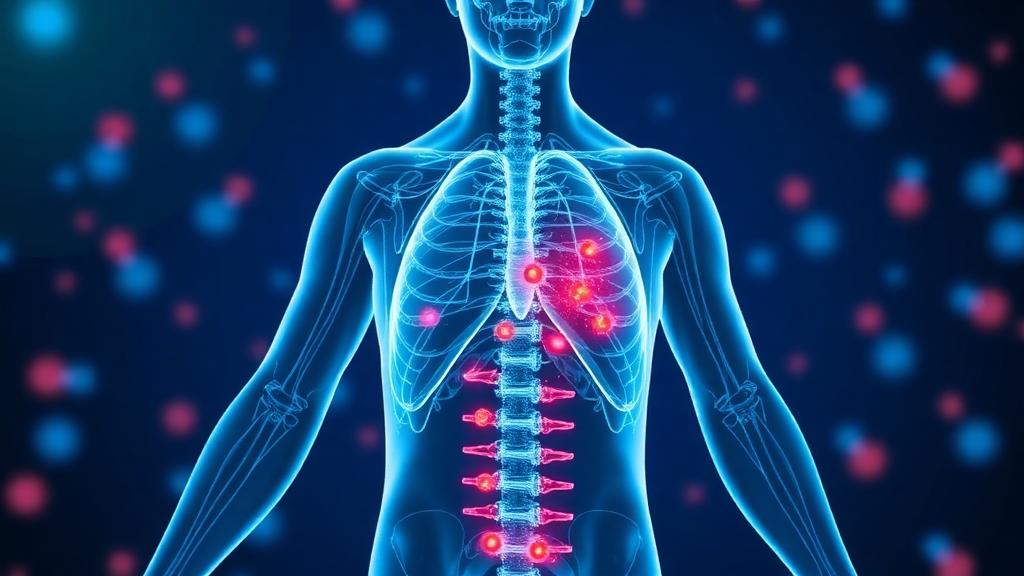
Cancer vaccines have delivered some of the most striking successes in modern immunotherapy, yet their results remain stubbornly unpredictable. Two patients with the same tumor type and stage can receive the identical vaccine and experience profoundly different outcomes, a variability that has frustrated oncologists and limited the broader clinical impact of mRNA and antigen-based platforms. Now, a team of researchers has built an ambitious computational tool designed to explain and predict that variability before a single dose is administered. Writing in Advanced Science, the group describes a whole-body, physiologically-based pharmacokinetic and pharmacodynamic model that simulates how a vaccine travels through the human body, how immune cells respond, and how a tumor fights back. The work, led by Mohammad R. Nikmaneshi with Timothy Padera and Lance Munn, was supported by the National Institutes of Health and represents one of the most comprehensive attempts yet to turn vaccination from an educated guess into a personalized prescription.
The core problem the model addresses is that vaccination is not simply a matter of injecting antigen and waiting for T cells to attack. Immune activation depends on a delicate choreography: antigen must meet immature antigen-presenting cells in peripheral tissue, and those activated cells must then travel through lymphatic vessels to lymph nodes, where they encounter naive T cells ready to be converted into tumor-killing effectors. Each step in this relay is shaped by anatomy. Blood flow rates, lymphatic drainage patterns, vessel permeability, and the specific lymph nodes draining a given tissue all influence whether the immune system mounts a vigorous response or a feeble one. Tumors complicate matters further by releasing suppressive factors such as TGF-beta, IL-10, VEGF, and lactic acid, which circulate systemically and can dampen immune priming in lymph nodes far from the tumor itself.
Existing computational models have captured fragments of this system, but the new framework is notable for its scope. It divides the body into interconnected compartments representing the heart, brain, lungs, liver, spleen, gastrointestinal tract, kidneys, breast tissue, skeletal muscle, bone, and a skin network split into six regions, each with its own draining lymph nodes. Arterial, venous, and lymphatic circulations link these compartments, and a modified version of Starling’s law governs fluid exchange between vessels and tissue. Crucially, each organ is paired with upstream and downstream lymph nodes arranged in series, each equipped with its own blood supply through high endothelial venules and its own afferent and efferent lymphatic pathways. This architecture allows the model to track where in the body T cell activation can actually occur, rather than assuming a single generic lymph node handles all priming.
At the cellular level, the model follows tumor cells, naive T cells born in the thymus, immature and activated antigen-presenting cells generated mainly in the bone marrow and skin, and effector T cells that proliferate in response to IL-2 and migrate into tumors. At the molecular level, it simulates antigen, inflammatory cytokines, IL-2, VEGF, and a lumped tumor-derived suppressive factor. Because quantitative data on individual suppressive pathways remain scarce, the researchers modeled tumor-induced immunosuppression as a single aggregate factor, an acknowledged simplification that keeps the system tractable while capturing its dominant effect. Naive T cells can only be activated in lymph nodes and the spleen, never in ordinary tissue, which makes the location and suppression state of those lymphoid organs decisive.
To quantify where activation is possible, the team introduced a concept called activation potential, essentially the probability that a given lymph node or the spleen can support T cell priming at a given moment. This probability decays exponentially as suppressive factor concentration rises. Simulations of a patient with a breast tumor vaccinated in the left arm revealed that suppressive factor accumulates fastest in the lymph nodes closest to the tumor’s drainage pathway, silencing them early. When suppressive factor production was raised by 40 percent, the window for effective vaccination in tumor-draining upstream lymph nodes shrank from roughly five days to just two or three. The model also showed that because all blood passes through the lungs, upstream lymph nodes draining the lungs are suppressed faster than their downstream counterparts, a subtle anatomical effect that simpler models would entirely miss.
The researchers then evaluated vaccination across every major organ and skin region, scoring outcomes with three metrics: activation site occupancy, which measures local activation in each lymphoid tissue; overall immune hotness, reflecting the total systemic expansion of effector T cells; and tumor immune hotness, the fraction of those cells that actually reach the tumor. The results were striking. For patients whose tumors produced high levels of suppressive factor, the farther the vaccination site sat from the tumor, the stronger the immune response, and injecting vaccine directly into the tumor performed poorly because tumor-draining lymph nodes were already saturated with suppressive signals. Conversely, patients with high tumor antigen production were far less sensitive to vaccination site, because tumor-released antigen was already activating lymph nodes throughout the body, making the vaccine a helpful supplement rather than the primary trigger.
Timing and route mattered as well. Intradermal and intramuscular vaccination produced similar systemic responses regardless of which skin region was used, suggesting both are viable delivery routes. In patients with high suppressive factor, early vaccination at day 7 favored internal sites in a specific order, with the gastrointestinal tract, spleen, bone, brain, and lungs outperforming the tumor, kidneys, and liver. Vaccination in the liver and kidneys proved ineffective across the board, because antigen injected there was rapidly cleared through vascular and lymphatic transport before antigen-presenting cells could take it up. For low-suppression patients with abundant antigen, the lungs yielded the strongest response. Notably, the model suggested that dual-site vaccination, combining an intratumoral dose with a distant one, could help patients whose tumors secrete high levels of suppressive factors.
Because running thousands of mechanistic simulations is computationally expensive, the team trained a random forest machine learning algorithm on the model’s outputs. The surrogate reproduced the mechanistic predictions with a correlation of 0.99, enabling rapid exploration of virtual patient populations and feature importance analysis. That analysis ranked tumor antigen level as the strongest predictor of tumor immune hotness, followed by suppressive factor level, vaccination timing, and vaccination site. When the researchers simulated vaccination on eight different days across five delivery routes for patients with unknown antigen and suppressive factor profiles, the estimated overall efficacy was highest for lung vaccination at 23.75 percent, followed by spleen at 23 percent, gastrointestinal and superficial routes near 21.5 to 21.75 percent, and intratumoral delivery last at 16 percent.
The model was calibrated and evaluated against experimental and clinical datasets covering antigen-dependent T cell activation, suppression by tumor-derived factors, and therapeutic vaccination responses, and the authors report good agreement with observations. They emphasize that the framework is a first step toward true digital twins of the immune system, which would require patient-specific parameterization from clinical and omics data, calibration against longitudinal measurements, and prospective validation. Still, the central message is already clear: identical vaccination protocols may produce substantially different immune responses across patients, and the optimal route and schedule depend on measurable features of each person’s tumor, particularly how much antigen and suppressive factor it releases. If validated prospectively, this kind of whole-body simulation could one day let oncologists test a vaccine virtually, choosing the site and timing most likely to turn a cold tumor hot before treatment ever begins.
Subject of Research: A physiologically-based pharmacokinetic/pharmacodynamic model for optimizing cancer vaccination strategies
Article Title: Optimizing Cancer Vaccinations Using a Physiologically‐Based Pharmacokinetic/Pharmacodynamic (PBPK/PD) Model
Article References: R. Nikmaneshi, M., P. Padera, T., & L. Munn, L. (2026). Optimizing Cancer Vaccinations Using a Physiologically‐Based Pharmacokinetic/Pharmacodynamic (PBPK/PD) Model. Advanced Science, Article e78044. https://doi.org/10.1002/advs.78044
Image Credits: AI Generated
DOI: 10.1002/advs.78044
Keywords: cancer vaccines, PBPK/PD modeling, immunotherapy, lymphatic system, T cell activation, tumor microenvironment, suppressive factors, personalized medicine, machine learning, systems immunology, lymph nodes, digital twin
News Source: Nathaniel Bowman. (October 4, 2026). Whole-Body Computer Model Aims to Pinpoint the Best Way to Vaccinate Each Cancer Patient. Scienmag.