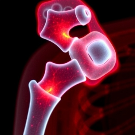
Bone health assessment has traditionally hinged on measuring bone mineral density (BMD), widely regarded as the definitive standard for fracture risk prediction. However, this well-established metric reveals a confounding paradox in patients with type 2 diabetes mellitus (T2DM). Despite presenting normal or even elevated BMD levels, these individuals exhibit markedly increased fracture susceptibility. This clinical contradiction highlights the inadequacy of mineral density alone in portraying true bone integrity, prompting a reassessment of what defines bone robustness. The critical insight emerging from current research underscores the significance of bone microarchitecture, organic matrix distribution, and osteocyte network integrity, all of which intricately contribute to mechanical stability beyond mere mineral content.
Capturing the subtle microstructural and compositional abnormalities that underlie diabetic bone fragility is notoriously challenging, particularly when these defects manifest at micrometer or nanometer scales. Conventional diagnostic modalities fall short here; standard histological techniques necessitate extensive chemical staining and dauntingly slow decalcification procedures. These methods not only consume precious time and labor but also irreversibly alter the native bone environment, impeding accurate pathological evaluation. Additionally, traditional optical microscopy often isolates single tissue components—be it collagen fibrils or mineral phases—resulting in disjointed, two-dimensional snapshots that fail to elucidate the complex three-dimensional biochemical interplay essential for comprehensive bone health understanding.
Addressing these limitations, a transformative approach employing multimodal nonlinear optical (NLO) microscopy has emerged as a cutting-edge solution for directly visualizing diabetic bone microdamage in situ, without exogenous labeling or destructive sample preparation. By harnessing intrinsic nonlinear optical phenomena and characteristic vibrational signatures within the bone matrix, this technique enables label-free “optical biopsies” that preserve the fragile microenvironment. The synergistic integration of stimulated Raman scattering (SRS), second harmonic generation (SHG), and two-photon excited fluorescence (TPEF) into a unified imaging platform allows for simultaneous mapping of proteins, lipids, and collagen fibers with ultrastructural precision. This multidimensional multimodal microscopy offers an unprecedented window into bone tissue composition and architecture, laying the groundwork for deep mechanistic insights into diabetes-related skeletal deterioration.
Recognizing that single-channel optical imaging often fails to capture comprehensive pathological nuances, a pioneering team led by Professor Ting Li from the Chinese Academy of Medical Sciences and Peking Union Medical College has synergized multimodal NLO microscopy with artificial intelligence (AI). Their collaboration with Beihang University and Shanghai East Hospital harnesses AI to deconvolute high-dimensional microscopic features hidden within multiplexed nonlinear optical images. Through sophisticated machine learning algorithms extracting spatial texture attributes across protein, autofluorescent metabolite, and phosphate channels, the team constructed robust classification models capable of distinguishing diabetic bone samples with an exceptional 93.56% accuracy. This performance significantly surpasses the approximate 70% accuracy of traditional single-channel optical diagnostics, demonstrating the potent advantage of multimodal data fusion coupled with advanced computational analysis.
Further enhancing pathological interpretability, the researchers employed explainable AI techniques to pinpoint a distinctive spatial degradation characteristic uniquely present in T2DM bone tissue. In healthy bone, protein distributions within osteocyte networks appear clustered with high contrast and intricate detail, reflecting preserved microstructural organization vital for mechanical function. Contrastingly, diabetic bone exhibits pronounced protein spatial homogenization—an aberrant uniformity and smoothness in protein optical texture indicative of osteocyte network disruption and loss of structural gradients. This novel optical marker, identified for the first time through AI-powered multimodal nonlinear imaging, provides a compelling biomarker of diabetic bone impairment, effectively labeling pathological alterations invisible to conventional imaging.
While these findings mark a revolutionary advance in osteoporosis and diabetic bone disease diagnostics, the translational journey towards clinical adoption necessitates further rigorous validation. Expanding clinical cohorts and incorporating multi-center studies will strengthen model generalizability across diverse patient populations. Complementary investigations integrating immunohistochemistry, proteomic profiling, and biomechanical assays will elucidate the molecular underpinnings of protein spatial homogenization, linking microscopic imaging phenotypes with biochemical and functional outcomes. Such multidisciplinary validation efforts promise to establish a solid foundation for routine clinical deployment of this transformative optical-AI diagnostic paradigm.
Beyond just diagnostic innovation, this research underscores the vast potential of combining label-free multimodal nonlinear optical microscopy with state-of-the-art artificial intelligence in biomedical science. This integrated approach transcends traditional imaging boundaries by facilitating nondestructive, high-resolution, molecularly specific visualization of complex tissue microenvironments. It opens unprecedented avenues for dissecting micro-pathological alterations across a broad spectrum of diseases, fostering new avenues for early detection, therapeutic monitoring, and mechanistic exploration of intricate biological systems.
The AI Theranostics Laboratory (AIT), established in 2018 and spearheading these developments, exemplifies interdisciplinary integration across optoelectronics, biomedical engineering, and neural computing. Their work seamlessly blends nonlinear optical sensing with deep learning to advance precision diagnostics and brain-machine interface technologies. With a prolific publication record and prestigious scientific accolades, AIT exemplifies how synergizing photonics and artificial intelligence can birth novel tools to untangle complex physiological phenomena, further accelerating translational impact.
This groundbreaking study, published in Opto-Electronic Advances (Impact Factor 22.4, 2024), depicts a major technological leap in visualizing diabetes-related skeletal compromise. The fusion of stimulated Raman scattering, second harmonic generation, and two-photon fluorescence modalities, coupled with AI-driven analysis, delivers a robust, high-accuracy diagnostic signature of T2DM-induced bone fragility. This integrative optical platform not only enhances diagnostic precision but also enriches biological understanding of osteocyte network degradation, representing a paradigm shift in bone pathology research.
Looking ahead, the next frontier lies in expanding this imaging and AI framework to other skeletal diseases and pathological contexts, potentially enabling broad-spectrum, label-free pathological phenotyping. Coupling this potent technology with emerging molecular biology and biomechanics could yield comprehensive disease models linking molecular perturbations to tissue mechanics. Such efforts will ultimately facilitate precision medicine strategies tailored to preserving bone integrity in diabetic and osteoporotic patients alike.
The promising success of merging multimodal nonlinear optical imaging with explainable AI heralds a new era in biomedical diagnostics that leverages intrinsic molecular contrasts and computational intelligence for unparalleled tissue microenvironment characterization. This innovative platform exemplifies the profound benefits of cross-disciplinary convergence, unlocking hidden biological information previously inaccessible to traditional methodologies. As this technology matures, it stands poised to substantially impact clinical diagnostics and research paradigms for complex bone disorders and beyond.
Subject of Research: Type 2 diabetes mellitus-induced bone fragility assessed by multimodal nonlinear optical imaging and AI
Article Title: AI-powered nonlinear optical imaging reveals protein spatial homogenization as an indicator of impaired bone quality in type 2 diabetes
News Publication Date: Not specified
Web References: http://dx.doi.org/10.29026/oea.2026.250312
References: Zhang BW, Pu JB, Hu T et al. AI-powered nonlinear optical imaging reveals protein spatial homogenization as an indicator of impaired bone quality in type 2 diabetes. Opto-Electron Adv 9, 250312 (2026). DOI: 10.29026/oea.2026.250312
Image Credits: oea
Keywords
label-free nonlinear optical imaging, type 2 diabetes mellitus, bone quality impairment, explainable AI, multimodal imaging integration, stimulated Raman scattering, second harmonic generation, two-photon excited fluorescence, osteocyte network, protein spatial homogenization
Tags: 3D biochemical bone matrix evaluationadvanced bone imaging techniquesAI-driven nonlinear optical imagingbone microarchitecture assessmentcollagen and mineral phase imagingdiabetic bone fragility detectionlimitations of bone mineral density in diabetesmicrometer-scale bone structural defectsnonlinear optical microscopy in bone researchosteocyte network integrity analysisprotein spatial homogenization in bonereduced bone quality in type 2 diabetes

