In the intricate ballet of human locomotion, vision serves as the critical navigator, continuously relaying environmental cues to the brain while intricately modulating motor decisions through sensorimotor integration. This multisensory interplay ensures stable and adaptive walking behavior under a variety of conditions. However, when visual input is compromised, the brain confronts a formidable challenge: how to sustain walking stability and fluidity despite diminished sensory information. Unraveling the neural compensatory mechanisms underlying this phenomenon holds immense promise, not only for elucidating fundamental brain function but also for pioneering novel therapeutic interventions to aid motor rehabilitation in individuals with visual impairments.
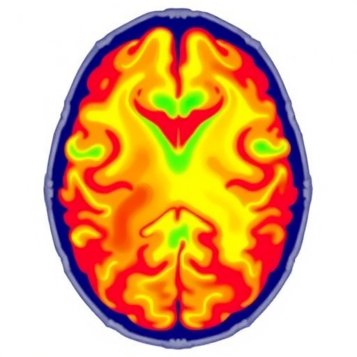
Recently, a groundbreaking investigation spearheaded by researchers at Peking University utilized Bangerter™ occlusion foils to simulate varying degrees of visual impairment in healthy young adults. This innovative approach enabled a controlled exploration of how reduced visual fidelity influences neural activity patterns linked to walking. Employing a sophisticated fusion of pattern-reversal visual evoked potentials (PR-VEPs) and resting-state functional magnetic resonance imaging (rs-fMRI), the study dissected the alterations in visual electrophysiology and intrinsic brain connectivity occurring after ambulation under different visual conditions. The research, published in the esteemed Chinese Medical Journal in March 2026, traverses the frontiers of neuroscience and rehabilitation medicine to decode the brain’s adaptive recalibration to sensory adversity.
The initial phase of the study confirmed that simulated visual occlusion markedly impairs the efficiency of signal processing along the visual pathway, as evidenced by diminished PR-VEP amplitudes. This validation underscores the robustness of the induced low-quality visual input model, establishing a reliable foundation for subsequent neuroimaging analyses. Such electrophysiological indexes are quintessential for gauging the immediate integrity of visual cortical processing and its downstream effects on sensorimotor networks.
Delving deeper, resting-state fMRI data elucidated dynamic changes in intrinsic brain activity following walking under contrasting visual states. Notably, the amplitude of low-frequency fluctuations (ALFF), a metric reflective of spontaneous neuronal activity, decreased significantly in the right paracentral lobule after walking with normal vision. Intriguingly, this diminution was partially reversed when walking was performed under visual occlusion, hinting at a compensatory neural rebound. This localized resurgence in activity may represent an adaptive upregulation aimed at maintaining sensorimotor integration and gait stability when visual cues are unreliable.
Beyond focal activity shifts, the study unveiled extensive modulation of functional connectivity across multiple sensorimotor circuits critical for locomotion. Specifically, enhanced synchrony was observed bilaterally between the calcarine cortex and middle temporal gyrus, regions traditionally implicated in primary visual and higher-order motion processing, respectively. This alignment suggests that visual-motor pathways remain engaged in locomotor control, even amidst diminished visual input, through heightened inter-regional communication.
Similarly, connectivity strengthened between the bilateral supplementary motor areas (SMA) and the right cuneus, an occipital region associated with early visual processing. This network appears pivotal for integrating motor planning with residual visual information during ambulation. The SMA’s role as a coordinator of voluntary movement likely facilitates anticipatory adjustments critical for navigating complex environments under sensory constraints.
Moreover, coupling between the bilateral precentral gyri—primary motor cortices—and the right cerebellar lobule VI was fortified. The cerebellum’s recognized function in fine motor coordination and error correction positions it as a central hub for refining gait adjustments when visual accuracy is compromised. Enhanced motor-cerebellar connectivity thus reflects a sensorimotor feedback loop vital for gait stability.
Most striking among connectivity changes was the marked fortification of functional interplay between the right precentral gyrus and right middle frontal gyrus exclusively under visual occlusion. This augmentation potentially serves as the brain’s central compensatory mechanism, recruiting executive and attentional networks to bolster motor control in the setting of unreliable visual input. The middle frontal gyrus, implicated in higher-order cognitive functions including working memory and attentional modulation, may facilitate the integration of multisensory information to compensate for visual deficits.
Collectively, the findings embody a dual-strategy neural adaptation: a rigid activation of fundamental sensorimotor pathways paired with targeted local connectivity enhancements within prefrontal and motor cortical regions. This balance ensures both the preservation of essential locomotor processes and the flexible recruitment of auxiliary circuits to offset sensory insufficiency. Such insights advance our understanding of brain plasticity and underscore the cerebellum and prefrontal cortex as crucial nodes in sensory compensation networks.
From a translational perspective, this research charts an innovative trajectory for motor rehabilitation paradigms tailored to low-vision populations. By harnessing the identified compensatory networks—particularly the augmented right precentral-middle frontal connectivity—therapeutic interventions can be designed to intensify functional integration within these circuits. Multimodal training approaches that synergize visual and somatosensory stimuli could potentiate these pathways, thereby enhancing gait stability and reducing fall risk.
Future avenues may encompass personalized neuromodulation strategies employing techniques like transcranial magnetic stimulation or neurofeedback to selectively enhance connectivity patterns revealed by this study. Furthermore, integrating wearable technology to provide augmented sensory feedback during locomotion could help reinforce adaptive brain activity patterns, providing real-time support to visually impaired individuals.
Professor Yingfang Ao of Peking University, leading this pioneering effort, emphasizes the transformative potential of these findings. With over two decades of expertise in sports medicine and exercise neuroscience, Professor Ao envisions a future where the neural mechanisms delineated here inform customized rehabilitation that operates at the intersection of brain function and behavioral therapy. This innovative fusion promises not only improved mobility outcomes but also greater autonomy and quality of life for those afflicted by visual impairments.
In summary, this comprehensive study reveals the cerebral adaptations that arise when walking occurs with compromised vision. By leveraging electrophysiological and neuroimaging modalities, it highlights the brain’s remarkable capacity to reconfigure functional connections, blending steadfast sensorimotor activations with strategic recruitment of prefrontal regions. Such discoveries not only deepen our fundamental neuroscientific knowledge but also catalyze the evolution of rehabilitation sciences, ushering in an era of precise, brain-guided intervention for sensory-motor deficits.
Subject of Research: People
Article Title: Resting-state functional magnetic resonance imaging study on the effects of visual status on walking-related brain functions in healthy young adults
News Publication Date: 20-Mar-2026
Web References:
Chinese Medical Journal Article DOI: 10.1097/CM9.0000000000004040
References:
DOI: 10.1097/CM9.0000000000004040
Image Credits: Chinese Medical Journal
Keywords
Neuroscience, Functional MRI, Visual Impairment, Sensorimotor Integration, Locomotion, Brain Connectivity, Rehabilitation, Visual Evoked Potentials, Motor Cortex, Cerebellum, Brain Plasticity, Multisensory Compensation
Tags: brain plasticity in visual deficitsmotor rehabilitation for visually impairedmultisensory processing in locomotionneural compensatory mechanisms walkingneural correlates of adaptive walkingpattern-reversal visual evoked potentialsresting-state fMRI brain connectivitysensorimotor integration in locomotionvisual impairment effects on walkingvisual modulation of gait stabilityvisual status and motor controlwalking-related brain activity

