
Headline: Revolutionizing the Diagnosis of Developmental Dysplasia of the Hip in Infants: A Focus on Ultrasound Evaluation
In recent years, developmental dysplasia of the hip (DDH) has emerged as a concerning orthopedic condition affecting infants worldwide. This developmental issue, characterized by improper formation of the hip joint, can lead to pain and functional limitations if not diagnosed and treated promptly. Thankfully, advancements in imaging technologies, particularly ultrasound, are reshaping the landscape of early diagnosis and management of DDH, enabling healthcare providers to make more informed decisions about treatment strategies.
The critical observation is that the hip joint’s normal development is crucial in the first few months of an infant’s life. The first six months are especially significant as the joint structures are still evolving. Traditional methods of diagnosing DDH, which often relied on clinical examination and X-rays, have their limitations—especially when it comes to assessing the hip’s dynamic nature in small, developing joints. As a result, the incorporation of ultrasound techniques in clinical practice has provided an invaluable alternative, offering real-time insights into the morphology and mobility of the hip joint.
.adsslot_RjfbdiTN4A{width:728px !important;height:90px !important;}
@media(max-width:1199px){ .adsslot_RjfbdiTN4A{width:468px !important;height:60px !important;}
}
@media(max-width:767px){ .adsslot_RjfbdiTN4A{width:320px !important;height:50px !important;}
}
ADVERTISEMENT
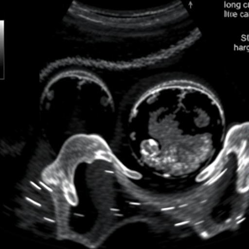
Ultrasound imaging, being non-invasive and devoid of ionizing radiation, has been embraced by pediatric specialists as the imaging modality of choice in assessing DDH. This modality enables professionals to visualize the hip joint in a manner that elucidates its anatomy and biomechanical behavior. This unprecedented visual access allows clinicians not only to identify abnormal joint configurations but also to monitor changes over time, facilitating a more personalized approach to treatment.
Given its advantages, the utilization of ultrasound in assessing DDH has seen a significant uptick. Clinicians now employ various ultrasound techniques to capture dynamic evaluations, which provide superior detail concerning the position and movement of the femoral head within the acetabulum. These insights prove crucial in determining the stability of the hip joint, as they help clinicians differentiate between stable and unstable configurations—a key factor in informing treatment options.
Furthermore, it is essential to highlight the role that training and experience play in obtaining accurate ultrasound results for DDH assessment. The nuances of infant hip ultrasounds require clinicians to be adept in both the technology of ultrasound and the pathology of hip development. Healthcare providers must possess a keen understanding of the normal and abnormal appearance of hip joints in varying developmental contexts to minimize the risk of misdiagnosis and improve patient outcomes.
The growing body of evidence surrounding the effectiveness of ultrasound in diagnosing DDH is underscored by ongoing research exploring its predictability and reliability. Recent studies have examined the correlation between ultrasound findings and long-term developmental outcomes, demonstrating promising results. These findings have potential implications for the clinical protocols of screening programs, particularly in high-risk populations where DDH incidence is disproportionately higher.
Moreover, the shift towards ultrasound is not just a clinical necessity; it is also a cost-effective strategy. Early diagnosis through ultrasound can lead to timely interventions, thereby curbing long-term complications associated with untreated DDH. Traditional treatment modalities often involve extensive postural management and surgery, which not only comes with substantial healthcare costs but also long-term impacts on the child’s quality of life. In contrast, early treatment initiated based on ultrasound assessments can mitigate many of these extensive treatment needs.
As health advocacy continues to push for improved neonatal screening practices, the integration of ultrasound technology in diagnosing DDH represents a critical step forward. The ability to screen infants in a comfortable, non-invasive environment fosters a strong trust relationship between healthcare providers and young families. Such early engagement can enhance compliance with follow-up care, ensuring that potential issues are addressed proactively.
However, as ultrasound technology expands its footprint in the assessment of DDH, it also brings to light challenges that need to be addressed. Standardization across various clinics and hospitals, accessibility of trained personnel, and variations in equipment quality can lead to disparities in diagnostic accuracy. Therefore, continued training and shared protocols among pediatric practitioners are essential, enabling consistency and reliability in using this imaging modality.
Overcoming these hurdles will likely involve collaborative efforts from medical societies, educational institutions, and healthcare organizations worldwide. Continued research is pivotal; finding uniform protocols can streamline training for clinicians and ensure that all practitioners, regardless of their geographical location, have access to the latest methods and interpretations regarding ultrasound images of hip joints.
In conclusion, the evaluation of developmental dysplasia of the hip using ultrasound represents a paradigm shift in pediatric healthcare. As technology continues to evolve, so too must our approaches to diagnosing and managing conditions like DDH. By harnessing the power of ultrasound, healthcare practitioners can ensure earlier diagnosis and intervention, ultimately leading to improved outcomes for children. This emphasizes the importance of integrative approaches that combine innovation, research, and clinical expertise to confront common pediatric challenges, thus making strides toward better health for the youngest patients.
Subject of Research: Developmental Dysplasia of the Hip
Article Title: Developmental dysplasia of the hip: ultrasound evaluation.
Article References:
Krauss, J., Straus Takahashi, M. & Samet, J. Developmental dysplasia of the hip: ultrasound evaluation.
Pediatr Radiol (2025). https://doi.org/10.1007/s00247-025-06334-y
Image Credits: AI Generated
DOI: https://doi.org/10.1007/s00247-025-06334-y
Keywords: Developmental Dysplasia of the Hip, Ultrasound, Pediatric Radiology, Early Diagnosis, Hip Joint Evaluation
Tags: advancements in ultrasound technology for infantsclinical management of hip dysplasiadevelopmental dysplasia of the hip in infantsdiagnosis of DDH using ultrasounddynamic assessment of hip joint developmentearly detection of hip joint abnormalitiesimaging advancements in infant orthopedicsimportance of early diagnosis in pediatric healthnon-invasive imaging techniques for hip assessmentpediatric orthopedic conditionstreatment strategies for developmental dysplasiaultrasound evaluation of hip dysplasia


