A groundbreaking study published in BMC Cancer has unveiled a novel positron emission tomography (PET) imaging biomarker that holds significant promise in predicting postoperative recurrence in lung adenocarcinoma (LUAD), the most common form of lung cancer. The research zeroes in on innovative PET parameters based on the spatial distribution of radiotracer uptake within tumors, providing a new frontier in precision oncology for operable lung cancer patients. This scientific advancement could potentially reshape postoperative surveillance strategies and personalized patient management.
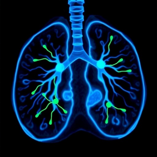
Lung adenocarcinoma, a subtype of non-small cell lung cancer, remains a formidable clinical challenge due to its tendency for postoperative recurrence even after surgical resection, which is currently the mainstay curative approach for early-stage disease. Predicting which patients are at higher risk for recurrence has remained elusive with conventional imaging biomarkers. Standard PET/CT parameters such as maximum standardized uptake value (SUVmax) commonly reflect tumor metabolism but fall short in predicting postoperative outcomes robustly. The emerging concept explored in this study is the spatial relationship of the metabolic “hot spot” — the point of highest radiotracer uptake — relative to key tumor anatomical landmarks.
The research team retrospectively analyzed data from 164 patients with surgically treated, pathologically confirmed stage IA–IIIA lung adenocarcinoma. All participants had undergone preoperative ^18F-Fluorodeoxyglucose PET/CT imaging, a powerful tool that maps glucose metabolism within tumors. Beyond conventional metabolic metrics, the researchers introduced and meticulously quantified two novel parameters: the normalized distance from the maximum uptake point (hot spot) to the tumor centroid, termed NHOCmax, and the normalized distance from the hot spot to the tumor perimeter, termed NHOPmax. These metrics effectively capture where within the tumor the metabolic peak is situated, normalized for tumor size, providing unique insights into tumor heterogeneity and aggressiveness.
Remarkably, the study found that NHOPmax, the distance from the highest glucose-avid point to the tumor’s outer edge, was the most potent predictor of postoperative recurrence and disease-free survival (DFS). It achieved an area under the curve (AUC) of 0.682 with an impressive sensitivity of 78.8%, outperforming traditional PET parameters in prognostic ability. This finding suggests that tumors with metabolic hot spots located closer to the perimeter rather than the center may confer a different biological behavior and risk profile, possibly reflecting invasive tumor fronts or areas of active proliferation.
Further statistical scrutiny demonstrated that NHOPmax was largely independent of other metabolic parameters like SUVmax, total lesion glycolysis (TLG), and metabolic tumor volume (MTV), indicating it conveys distinctive prognostic information. In both univariate and multivariate logistic regression analyses, NHOPmax showed a robust inverse association with postoperative recurrence risk, symbolizing that higher NHOPmax values — meaning the hot spot is positioned further from the perimeter — corresponded to superior patient outcomes.
Survival analysis added compelling weight to these observations, establishing NHOPmax as an independent predictor of disease-free survival. Patients with NHOPmax values exceeding the threshold of 0.43 experienced significantly longer DFS, underscoring the clinical utility of this novel imaging biomarker in stratifying recurrence risk. Integrating NHOPmax into postsurgical follow-up protocols could enable clinicians to tailor adjuvant therapies more precisely and optimize patient counseling.
The introduction of spatial PET parameters like NHOPmax transcends the traditional reliance on metabolic intensity alone. This paradigm shift emphasizes tumor microenvironment organization and heterogeneity as critical facets influencing cancer progression. By quantifying the positional metabolic gradients within tumors, clinicians could gain refined insights into tumor biology and behavior, potentially applicable beyond lung adenocarcinoma to other solid tumors.
Such an imaging biomarker dovetails seamlessly with the growing field of radiomics, where complex image features are computationally extracted and leveraged for clinical predictions. NHOPmax exemplifies a clinically actionable radiomic feature distilled from widely accessible PET/CT scans, enhancing translational value. Future research integrating NHOPmax with molecular and genomic tumor profiles could unlock synergistic prognostic models, propelling the era of precision oncology forward.
This study’s findings are especially poignant in the context of stage IA–IIIA lung adenocarcinoma, where surgical resection yields curative potential but recurrence risk remains a pressing concern. Current prognostic tools, including tumor-node-metastasis (TNM) staging, lack granularity in identifying which resected patients harbor micrometastatic disease or aggressive tumor phenotypes. NHOPmax adds a layer of nuanced, noninvasive risk stratification that could redefine postoperative monitoring intensity and therapeutic decision-making.
Moreover, the ease of calculating NHOPmax from routine ^18F-FDG PET/CT scans elevates its clinical feasibility. Since PET/CT imaging is standard for lung cancer staging, implementing NHOPmax quantification would require minimal alterations to imaging protocols, facilitating seamless adoption. This methodology also circumvents the need for invasive tissue sampling or complex molecular assays, democratizing risk assessment in diverse clinical settings.
While the current study is retrospective and single-institutional, it paves the way for prospective multicenter trials validating NHOPmax’s prognostic prowess. Evaluating its predictive capacity in conjunction with novel systemic therapies such as immunotherapy or targeted agents could further elucidate its role in evolving lung cancer treatment landscapes. Additionally, refining computational algorithms for automated NHOPmax measurement may enhance reproducibility and expedite clinical workflows.
In essence, this research pioneers a new dimension in oncologic imaging biomarkers by leveraging the spatial metabolic architecture of tumors. NHOPmax emerges not just as a statistical predictor, but as a window into the biological complexity underpinning tumor aggressiveness and recurrence. By translating this insight into clinical practice, oncologists may soon wield a powerful tool to preempt postoperative relapse and personalize patient care.
Together, these advancements highlight the transformative potential of enhancing PET imaging metrics beyond conventional parameters. The nuanced evaluation of glucose metabolism topography within lung adenocarcinoma introduces a critical step forward in precision diagnostics, prognostics, and therapeutics. As medicine gravitates towards individualized approaches, such innovative imaging biomarkers will undoubtedly play a pivotal role in shaping future lung cancer management strategies.
The implications extend beyond recurrence prediction: NHOPmax and similar spatial biomarkers might serve as early surrogate endpoints in clinical trials or as markers to select patients for intensified adjuvant therapies. They could also stimulate biologic investigations into the mechanisms driving differential metabolic distribution, unveiling novel targets to thwart invasion and metastasis.
In conclusion, the study’s identification of NHOPmax from ^18F-FDG PET/CT scans as a robust, independent predictor of postoperative recurrence in lung adenocarcinoma represents a major stride in oncologic imaging and prognosis. Its incorporation into clinical workflows promises to refine patient stratification, inform treatment decisions, and ultimately improve survival outcomes in this challenging malignancy. As the oncology community embraces increasingly sophisticated imaging analytics, such breakthroughs underscore the synergistic power of technology and clinical science in confronting cancer’s complexities.
Subject of Research: Predictive PET imaging biomarkers for postoperative recurrence in lung adenocarcinoma
Article Title: Novel PET imaging biomarkers as predictors of postoperative recurrence in lung adenocarcinoma
Article References:
Zheng, C., Miao, J., Xu, L. et al. Novel PET imaging biomarkers as predictors of postoperative recurrence in lung adenocarcinoma. BMC Cancer 25, 874 (2025). https://doi.org/10.1186/s12885-025-14263-0
Image Credits: Scienmag.com
DOI: https://doi.org/10.1186/s12885-025-14263-0
Tags: BMC Cancer study findingslung adenocarcinoma postoperative outcomeslung cancer recurrence predictionmetabolic hotspots in tumorsnon-small cell lung cancer challengespersonalized patient management in cancerPET imaging biomarkerspostoperative surveillance strategiesprecision oncology innovationsspatial distribution of radiotracer uptakesurgical resection of lung cancerSUVmax limitations in lung cancer
