According to a study published recently in the advanced edition of the journal Bone whose authors are members of the BCN MedTech research group; the company Galgo Medical, a UPF spin-off, the QUAES-UPF Chair, and the CETIR radiological center
Credit: UPF
Osteoporosis is a skeletal disease in which there is a decrease in bone mass density. The bones become more porous and fragile making them more susceptible to fracture. This disease reduces bone density and weakens the bone. The weakening of the bone increases the risk of fracture. Among all possible osteoporotic fractures, hip fractures are a major problem in Western countries. In fact, it is estimated that they affect one-third of women and a fifth of men. Hip fracture reduces mobility, quality of life and may even increase mortality in women and men. As such, the prediction of osteoporotic hip fractures is very important in terms of quality of life and life expectancy.
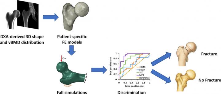
The main goal of a recent study published in the journal Bone was to find biomechanical criteria for the discrimination of the risk of hip fractures by exploring DXA-based 3D models and specific finite element simulations for patients in vivo. Carlos Ruiz Wills, first author of the study, and Jérôme Noailly, study coordinator, both members of the Biomechanics and Mechanobiology laboratory at the BCN MedTech Research Unit explain:
“This research has demonstrated that the modelling and biomechanical simulation of the bone by means of finite element-based 3D reconstructions of conventional bone densitometry provide descriptors of the internal tissue mechanics that go beyond the traditionally explored bone density when it comes to discriminating the risk of osteoporotic fracture of the proximal femur (hip fracture)”.
The finite elements method is a numerical calculation method widely used in simulations of complex physical and biological systems that enables solving differential equations associated with physical problems on complicated geometries.
This study combines the competencies of the Biomechanics and Mechanobiology laboratory in computational biomechanics and the competencies of the SiMBIOSys group at BCN MedTech, directed by Miguel A. Gonzalez Ballester (ICREA), in biomedical image analysis. Such synergy illustrates new trends in exploiting the potential of models and simulations to improve patient diagnosis, explain Jérôme Noailly and Miguel A. González Ballester: “On the one hand, advanced image analysis provides a customized framework of modelling and augmented reality, by integrating the morphology and the densities of the patients’ bones into virtual models. Moreover, the conversion of these models into finite element models capable of integrating equations of the bone’s mechanical behaviour in the event of external mechanical events, such as a fall, enables calculating descriptors that uniquely integrate crossed effects between bone quality, particular bone morphology and external mechanical forces that often depend on the weight and height of the patient”.
“The 3D models obtained in this study come from the reconstruction of flat 2D image densitometry (DXA, dual energy X-ray absorptiometry) using the 3D Shaper software developed by Galgo Medical, a UPF spin-off technology company”, comments Luis Miguel del Rio, a radiologist at the CETIR radiology centre (Ascires Group) and a contributor to the study. The potential of this biomedical technology comes from the fact that the simulations obtained by this research include three-dimensional interaction between bone density, the geometry of the femur and external mechanical loads, which cannot be measured in a patient.
The results obtained in this study show a power of discrimination usually greater than 80% compared to the calculation of hip fracture risk following a fall by the patient. In addition, in discriminating the risk of fracture, the simulations have allowed the authors to grant relative importance to the stress state of the trabecular bone, compared to the stress state of the cortical bone.
###
The study, published recently in the advanced edition of the journal Bone is the result of active collaboration by Carlos Ruiz Wills, first author; Andy Luis Olivares, Simone Tassani, Mario Ceresa, Veronika Zimmers, Miguel A. Gonzalez Ballester (ICREA), and Jérôme Noailly, study coordinator, all members of the BCN MedTech Research Unit of the Department of Information and Communication Technologies (DTIC) at UPF, the company Galgo Medical (UPF spin-off), and the CETIR radiological centre (Ascires Group). This scientific and technological cooperation between private and public centres has been possible thanks to such instruments as the QUAES-UPF Chair and the project Collaboration Challenges BioDXA, with the aim of promoting research and knowledge transfer in the field of computational technologies applied to health.
Media Contact
Nuria Pérez
[email protected]
Related Journal Article
http://dx.


