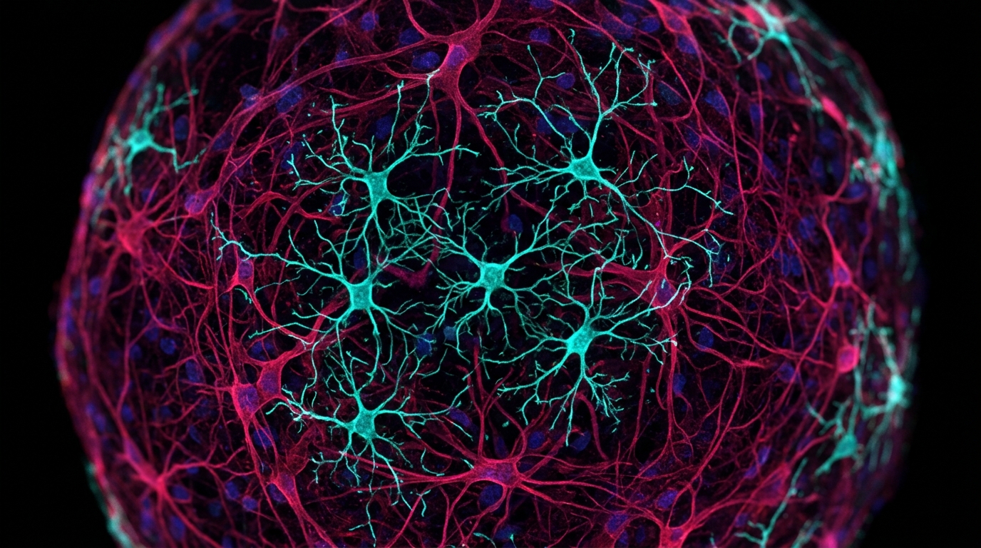
Researchers have demonstrated that primary rat cortical microtissues containing innate microglia undergo pronounced changes in cellular morphology, surveillance area, motility, and injury response across distinct stages of tissue maturation. In a study published in the Journal of Neuroinflammation, investigators Alexander Del Toro, Angelina Clark, Kaylen Aguilar, Alexander Bautista, Nathan Ashby, and Diane Hoffman-Kim showed that these three-dimensional neural multicellular assemblies enable real-time tracking of immune cell dynamics as the culture matures from early to late developmental phases.
Microglia represent the primary immune component of the central nervous system, displaying dynamic and multifaceted reactions to both tissue injury and disease states. Throughout both developing and mature nervous tissue, these immunocompetent cells continuously survey their surrounding microenvironment to maintain homeostasis and protect against potential threats. However, researchers have faced an ongoing gap in experimental three-dimensional in vitro models that successfully incorporate microglia while reproducing both developmental characteristics and mature functional hallmarks observed in living tissue.
To address this experimental challenge, the research team developed a three-dimensional multicellular cortical microtissue platform derived directly from primary rat cortical cells. Crucial to their investigation was the optimization of a specialized within-micromold imaging technique. This methodological approach permits direct live imaging of microglia without requiring the microtissues to be removed or transferred from their culturing environment, thereby preserving tissue conditions while enabling continuous observation across early, middle, and late maturation stages.
Through this live-imaging method, the researchers confirmed that resting microglia exhibit characteristic baseline surveillance within the three-dimensional microenvironment. The observed surveillance behavior is distinguished by relatively stationary cell somas operating alongside highly dynamic cell processes. These cellular extensions continuously extend into and retract from the surrounding matrix, allowing the stationary cell bodies to monitor adjacent areas actively without requiring immediate whole-cell translocation during baseline conditions.
The investigators also subjected the cortical microtissues to inflammatory stimuli to evaluate how the resident immune cells react to physiological threats. Following proinflammatory challenges, the microglia actively engulfed lipopolysaccharide particles within the three-dimensional environment. This phagocytic uptake was accompanied by noticeable dynamic shifts in the cells’ motility patterns, demonstrating that inflammatory exposure directly alters how these cells move and interact with their surroundings.
In addition to chemical challenges, the team evaluated the immune response to physical trauma by introducing targeted laser-induced tissue damage within the microtissues. The resident microglia responded rapidly to the focal injury through three coordinated behaviors: the extension of cell processes toward the lesion, whole-cell displacement across the matrix, and targeted local recruitment around the site of damage. Importantly, the researchers observed that the age of the microtissue in culture strongly influences both baseline motility profiles and directed motility responses toward injury.
Collectively, the experimental findings establish that microglia within a three-dimensional cortical microenvironment display substantial modifications in morphology, surveillance territory, motility behaviors, and injury responsiveness across microtissue maturation. The authors conclude that these primary rat cortical microtissues provide a valuable in vitro platform suitable for developmental investigations of microglia as well as experimental studies of central nervous system inflammation associated with brain injuries, infections, and neurological diseases.
Subject of Research: Real-time imaging of microglia dynamics in 3D rat cortical microtissues
Article Title: 3D cortical microtissue with innate microglia for studying real-time cell behavior across maturation and inflammatory response
Article References: Del Toro, A., Clark, A., Aguilar, K., Bautista, A., Ashby, N., & Hoffman-Kim, D. (2026). 3D cortical microtissue with innate microglia for studying real-time cell behavior across maturation and inflammatory response. Journal of Neuroinflammation. https://doi.org/10.1186/s12974-026-04039-z
Image Credits: AI Generated
DOI: 10.1186/s12974-026-04039-z
Keywords: microglia, cortical microtissues, 3D cell culture, in vitro models, primary rat cortical cells, live cell imaging, within-micromold imaging, baseline surveillance, cell motility, lipopolysaccharide, laser-induced damage, neuroinflammation, central nervous system, tissue maturation, process extension, tissue homeostasis
News Source: Ophelia Keating. (November 19, 2026). 3D Rat Cortical Microtissues Reveal Changing Microglia Dynamics During Tissue Maturation. Scienmag.