Credit: Tel Aviv University
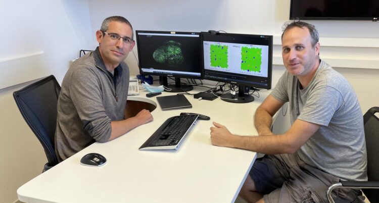
Using Zebrafish, researchers from the School of Neurobiology, Biochemistry and Biophysics at the Faculty of Life Sciences of Tel Aviv University have developed an advanced simulation of a key process in the brain – the activation of the stem cells responsible for generating neurons. The simulation revealed that the process, which until today was considered to be random, is in fact coordinated, thereby ensuring the normal production of neurons in the brain. According to the researchers, their findings add another layer of understanding to brain development, as well as to the study of brain damage and diseases, such as brain cancer and degenerative diseases such as Alzheimer’s and Parkinson’s. The simulation was based on the imaging of Zebrafish brains using innovative technology.
The study was led by Prof. David Sprinzak and research student Udi Binshtok of the School of Neurobiology, Biochemistry and Biophysics at the George S. Wise Faculty of Life Sciences, and Prof. Laure Bally-Cuif and her team from Institut Pasteur and The French National Centre for Scientific Research (CNRS). The article was published in the journal Cell Stem Cell.
Prof. Sprinzak explains that in the brains of all vertebrates, including humans, there are stem cells whose job is to produce new neurons (unlike embryonic stem cells, which can differentiate into all existing types of cells in the body, these stem cells can only produce neurons or additional stem cells).
In humans, these cells are active mainly in childhood, while the brain is developing. In an adult they are usually dormant, and spring to action only rarely and at specific locations, for example to form normal memory episodes.
Prof. Sprinzak adds that in degenerative brain diseases, such as Parkinson’s and Alzheimer’s, the process goes awry and there is a decrease in the number of stem cells in the brain and in their proliferation rate. In cases of brain cancer, in contrast, stem cells may become over-activated and proliferate uncontrollably, or different cells in the brain may turn back into stem cells, ensuring tumor persistence.
According to the researchers, the current study sought to examine the mechanism that activates the stem cells in the brain initiating cell division: “Until now, the activation was considered to be random, and we wanted to see if there was actually any order to it, and whether the activation of one stem cell affects others,” says Prof. Sprinzak. “To this end, we conducted research on Zebrafish, in which the laboratory at the Pasteur Institute in France specializes. Zebrafish were chosen because the stem cells in their brains are similar to those of mammals, but are much more numerous, so new neurons in their brains are constantly added.”
The study used advanced imaging technology that allowed the fish’s brain to be filmed while it is alive and active. The researchers filmed the fish every three days for a few weeks, creating a sequence of images that showed the process of neural stem cell activation and neuronal generation in the brain.
This data was transferred to Prof. Sprinzak’s laboratory at Tel Aviv University, which analyzed it and developed a computational simulation of the process of stem cell activation in the brain.
Prof. Sprinzak: “Our main discovery is that, contrary to what scientists have believed to date, the process is not random. The new neurons are formed in a coordinated manner. The computational model clearly showed that a stem cell undergoing activation does not allow the cells adjacent to it to undergo a similar process for several days. In this way, it ensures that the neurons produced are arranged in the brain properly. This finding is very important for understanding the normal development and structure of the brain.”
Prof. Sprinzak concludes: “Our research adds an important layer of understanding to brain development. It may help in the development of future treatments based on using neural stem cells, such as for brain damage and neurodegenerative diseases like Alzheimer’s and Parkinson’s where stem cells could be activated to produce new neurons. Furthermore, it advances our understanding of the possible mechanisms by which cancer develops in the brain as a possible infrastructure for treatments that could neutralize the cancer process.”
###
Media Contact
Noga Shahar
[email protected]
Related Journal Article
http://dx.


