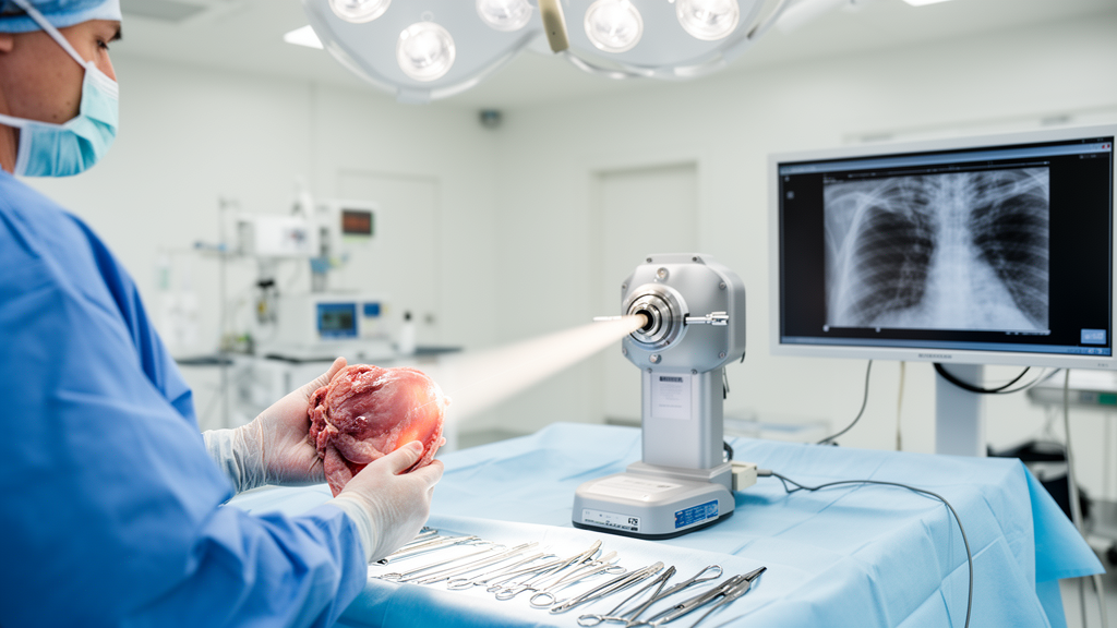
Every year, thousands of patients with early-stage breast cancer undergo surgery that removes the tumor along with a rim of apparently healthy tissue. Yet the operation carries an uncomfortable gamble: whether the surgeon has truly cut beyond the cancer. The answer usually arrives weeks later, when a pathologist examines the removed tissue under a microscope. If cancer cells are found at the edge of the specimen, the patient must return to the operating room. Now, a review published in Frontiers in Medicine argues that a technology borrowed from physics could end this waiting period by allowing surgeons to see cancer in real time, while the patient is still on the table.
The stakes are far from trivial. Reoperations expose patients to additional anesthesia, pain, and psychological distress, and they are associated with poorer cosmetic outcomes and diminished quality of life. They also carry substantial costs for healthcare systems. The review, led by Alessandro Olivo and colleagues at University College London, points out that the incidence of positive margins in breast conserving surgery remains high enough across hospitals to make real-time assessment a pressing clinical need. The core problem, however, is that cancer in soft tissue is notoriously difficult to see with conventional X-ray imaging, which excels at visualizing bone but struggles to distinguish one type of soft tissue from another.
The technology at the heart of the review is called X-ray phase contrast imaging. Unlike conventional X-rays, which measure how much radiation is absorbed as it passes through tissue, phase contrast imaging detects how the X-ray wave is subtly bent, or refracted, as it travels through different materials. Because this bending depends on the refractive index of the tissue, which varies far more between soft tissues than their absorption does, phase contrast images can reveal boundaries between tissues that would otherwise remain invisible. The effect is so sensitive that it can distinguish cancerous from healthy tissue in organs such as the breast, liver, and lung.
For decades, phase contrast imaging was confined to large facilities such as synchrotrons, which produce X-ray beams of exceptional quality but are impractical for clinical use. The review describes how, over the past two decades, physicists have developed methods that work with conventional X-ray sources. One approach, called Talbot-Lau interferometry, uses gratings to create interference patterns that reveal phase shifts. Another, known as edge illumination, masks the X-ray beam so that only its edges pass through the sample, amplifying the refraction effect. A third method, called propagation-based imaging, simply allows the X-ray wave to travel a distance after passing through the tissue, so that refraction becomes visible as a change in intensity.
The review highlights a study in which researchers used edge illumination to image breast tissue specimens with a compact setup. The results were striking: the images revealed cancerous lesions with a clarity comparable to that achieved at synchrotrons. But there was a catch. The scans took between five and ninety hours to complete, far too slow for clinical practice. The bottleneck lay in a procedure called phase stepping, in which one of the gratings must be moved in small increments for every angular position of the sample, multiplying the scan time.
The breakthrough came when Olivo and colleagues realized that edge illumination could be combined with a single-shot phase retrieval method originally developed for propagation-based imaging. Instead of stepping the grating through multiple positions, the new approach retrieves the phase information from a single exposure. The result was a dramatic acceleration: scans that once took hours could be completed in minutes. In a follow-up study, the researchers demonstrated that they could image an entire breast specimen in under ten minutes, a time compatible with surgical workflows.
The clinical implications were tested in a study that scanned breast tissue specimens from patients undergoing surgery. The images were assessed by radiologists who were blinded to the pathological findings. The results showed that the method could detect cancer with a sensitivity 2.6 times higher than conventional X-ray imaging, which is the current standard for specimen radiography. The images also revealed the extent of the tumor, including whether cancer cells reached the edge of the specimen, information that would allow surgeons to remove additional tissue immediately rather than waiting for the pathology report.
The review also describes how the technology could be applied beyond breast surgery. Phase contrast imaging has been used to visualize liver tumors and lung lesions in preclinical studies, suggesting that the method could assist surgeons in a range of operations where soft tissue boundaries are critical. The researchers note that the compact setups required for clinical use are becoming increasingly available, and that the technology is compatible with the imaging standards used in hospitals.
Challenges remain before the technology can be adopted in operating rooms. The review points out that metallic markers, which surgeons sometimes place in tissue, can create artifacts that obscure the images, although methods to correct for these artifacts have been developed. The researchers also note that the sensitivity of the method depends on the quality of the X-ray source, and that higher-power sources, which produce clearer images, are more expensive. The cost of the gratings used in some setups, currently between ten and twenty thousand dollars, could fall if they were mass-produced rather than custom-built.
Despite these hurdles, the review argues that the technology has reached a point where clinical translation is realistic. The ability to see cancer in real time, while the patient is still on the operating table, could transform surgery for breast cancer and beyond. The researchers conclude that the convergence of compact X-ray sources, sensitive phase contrast methods, and rapid image reconstruction has created a window of opportunity to bring this physics-based technology from the laboratory to the operating room.
Subject of Research: Physics
Article Title: X-ray phase contrast for intra-operative specimen imaging in breast conserving surgery and other areas
Article References: Olivo, A., & Leff, D. R. (2025). X-ray phase contrast for intra-operative specimen imaging in breast conserving surgery and other areas. Frontiers in Medical Engineering, 3, Article 1608247. https://doi.org/10.3389/fmede.2025.1608247
Image Credits: AI Generated
DOI: 10.3389/fmede.2025.1608247
Keywords: X-ray phase contrast imaging, breast conserving surgery, real-time cancer detection, specimen radiography, operating room technology, soft tissue imaging, clinical translation, X-ray, phase, contrast, intra-operative, specimen
News Source: Ophelia Keating. (October 17, 2026). X-ray phase contrast for intra-operative specimen imaging in breast conserving surgery and other areas. Scienmag.