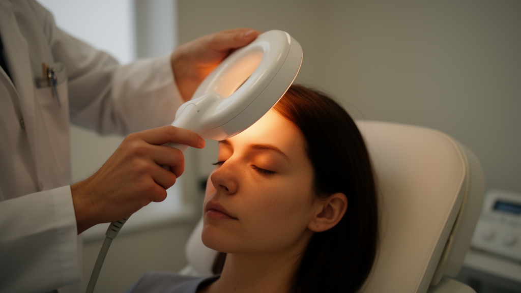
Transcranial magnetic stimulation, or TMS, has quietly become one of the most versatile tools in modern neuroscience. By passing a rapidly changing current through an insulated coil held against the scalp, clinicians can generate a magnetic field that passes unattenuated through the skull and induces an electric field in the outer few millimeters of the cortex, temporarily exciting or suppressing neural circuits without surgery. The technique is already an approved treatment for depression and is increasingly used to manage chronic pain. Yet a sweeping new scoping review published in Frontiers in Neuroscience suggests that one of the most consequential settings in the entire procedure — the angle at which the coil is held — has been treated far too casually, especially when the goal is pain relief.
The review, conducted by Armina Saadatkhah and Albert Leung of the University of California, San Diego, followed PRISMA-ScR guidelines and searched seven databases spanning publications from 1992 to 2025. After screening more than 1,200 records, the authors identified 24 studies related to the primary motor cortex, comprising 8 computational modeling studies and 16 human experiments, along with just three studies involving the dorsolateral prefrontal cortex, a favored target for depression and pain protocols. Risk of bias in the 18 human studies was assessed with the ROBINS-I V2 instrument. What emerged was a picture of a field that has rigorously optimized coil angle for moving muscles but almost entirely ignored it for relieving pain.
The technical heart of the matter lies in the induced electric field. The figure-of-eight coil most commonly used clinically focuses stimulation at the junction of gray and white matter, where corticospinal neurons can be depolarized. But the direction of that field relative to cortical anatomy matters enormously. Computational models built from MRI-based head models, using methods such as the finite element and boundary element approaches, consistently show that the strongest field — and the lowest neuronal activation threshold — occurs when the induced field runs perpendicular to the local sulcal wall and parallel to the principal axis of the underlying cortical columns. For the hand area of the motor cortex, that geometry happens to correspond to roughly 45 degrees in the posterior-to-anterior direction, the famous PA45° convention referenced to the interhemispheric fissure.
That 45-degree rule, it turns out, is not arbitrary but also not universal. Landmark experiments from the early 1990s by Brasil-Neto and colleagues and by Mills and colleagues found maximal motor evoked potentials when the induced current flowed diagonally across the central sulcus, and decades of subsequent work have confirmed the convergence. But the review emphasizes that PA45° is really a statement about the hand representation specifically. One of the largest computational efforts, by Gomez-Tames and colleagues, ran 518,616 simulated stimulation scenarios across 18 individualized head models and found that optimal orientation tracks local gyral anatomy with striking fidelity — but shifts away from 45 degrees elsewhere in the brain. Lee and colleagues added that individual anatomy matters too: brain-scalp distance and cerebrospinal fluid thickness together accounted for about 80 percent of the variability in peak field strength, producing nearly threefold differences in stimulation intensity between people receiving identical protocols.
The deviations become dramatic outside the hand knob. For lower-limb muscles such as the tibialis anterior, experimental studies found peak responses near PA90°, perpendicular to the midline rather than oblique to it, and the two orientations even produced qualitatively different intracortical inhibition profiles depending on whether the muscle was at rest or contracting. At the pre-supplementary motor area, the most selective activation in EEG recordings occurred at PA0°, while rotating to PA90° increased overall potency at the cost of spreading activation into adjacent motor cortex. Even within the hand area, individual optima can vary by as much as plus or minus 30 degrees, and the shape of a person’s hand knob — inverted-omega versus epsilon — can determine whether PA45° or PA90° works better. A Bayesian closed-loop algorithm called KG-BOOST found optimal orientations within about 5 to 7 degrees of PA45° on average but with meaningful individual spread, using 40 percent fewer pulses than a dense grid search.
The dorsolateral prefrontal cortex presents an even thornier problem. Unlike the motor cortex, the prefrontal target has no real-time physiological readout analogous to the motor evoked potential, so most clinical protocols simply borrow the motor-derived PA45° default. Only one of the three eligible DLPFC studies actually manipulated orientation: Thomson and colleagues showed with near-infrared spectroscopy that hemodynamic responses were largest at PA45°. A combined modeling and experimental study in Alzheimer’s patients and healthy controls found that functional targets showed 2.63-fold greater spatial dispersion than anatomical ones and that aligning the coil perpendicular to the functional-anatomical axis produced roughly 70 percent stronger stimulation at the functional target relative to surrounding tissue. On the basis of three studies, the review concludes, the 45° convention at the DLPFC is best regarded as an untested default rather than a validated optimum.
Then comes the review’s central and most striking finding: a void. Analgesic rTMS is thought to work through entirely different circuitry than the corticospinal system that motor evoked potentials measure. At the motor cortex, stimulation is believed to engage descending inhibitory pathways through the thalamus, periaqueductal gray, and rostroventromedial medulla, while prefrontal stimulation is thought to modulate the cognitive and affective appraisal of pain through prefrontal-limbic connectivity. A coil angle that maximizes corticospinal output is therefore not guaranteed to optimally engage the networks responsible for pain relief. Yet across every database searched, no study ever compared coil orientations for therapeutic analgesic outcomes. The 33 double-blind controlled trials synthesized in a prior review all used a standardized near-PA45° orientation without testing alternatives, meaning the contribution of coil angle to analgesic efficacy in every existing pain trial is confounded and unknown.
The single study that touched pain at all did so indirectly. Tani and colleagues measured pain thresholds during single-pulse TMS over Broca’s area and the motor hand knob across seven orientations, finding that thresholds varied significantly with angle at Broca’s area but not at the motor cortex — evidence that even the discomfort caused by the pulse itself can be orientation-dependent. But that study examined healthy subjects at non-clinical targets, leaving the primary stimulation sites for therapeutic pain rTMS entirely uncharacterized. The authors argue this negative finding is itself a substantive result: it defines a specific, testable research question rather than a vague under-researched area.
The review also offers practical guidance that does not require high-end equipment. A tiered workflow proposes using target-specific heuristic angles as a baseline — roughly PA45° for the hand area, PA0° for the pre-supplementary motor area, and PA90° or PA270° for lower-limb targets — then empirically testing a few angles around that heuristic to find the lowest motor threshold when physiology permits, and finally deploying individualized MRI-based electric-field modeling at centers where precision matters most. Applying the hand-area convention to a lower-limb target, the authors stress, is not a neutral default but a predictable and avoidable error. What remains untested is whether any of these tiers actually improves clinical outcomes, a question that will require pain trials reporting coil orientation alongside standardized measures such as quantitative sensory testing and numerical pain scores.
For a field increasingly betting on neuromodulation to treat chronic pain, the message is unusually actionable. Unlike cortical anatomy or network connectivity, coil orientation costs nothing to change during any stimulation session. The authors caution that it is one addressable source of variability among many — target definition, coil-to-cortex distance, intensity, pulse waveform, and dose all contribute — and none has been systematically compared against orientation for relative influence on outcome. But as computational dosimetry grows more sophisticated and individualized head models become routine, the humble rotation of a coil by a few dozen degrees may prove to be one of the cheapest levers available for making brain stimulation genuinely personal. Until orientation-controlled pain trials exist, however, the angle that best relieves suffering remains an open — and now precisely framed — question.
Subject of Research: Optimal coil orientation in transcranial magnetic stimulation and its untested role in analgesic outcomes
Article Title: TMS coil orientation: from computational modeling to experimental evidence and analgesic considerations
Article References: Saadatkhah, A., & Leung, A. (2026). TMS coil orientation: from computational modeling to experimental evidence and analgesic considerations. Frontiers in Neuroscience, 20, Article 1938946. https://doi.org/10.3389/fnins.2026.1938946
Image Credits: AI Generated
DOI: 10.3389/fnins.2026.1938946
Keywords: transcranial magnetic stimulation, coil orientation, electric field modeling, primary motor cortex, dorsolateral prefrontal cortex, rTMS, pain relief, neuromodulation, cortical anatomy, motor evoked potentials, scoping review, brain stimulation
News Source: Cassandra Pierce. (October 19, 2026). Why the Angle of a Brain-Stimulation Coil Could Make or Break Pain Relief. Scienmag.