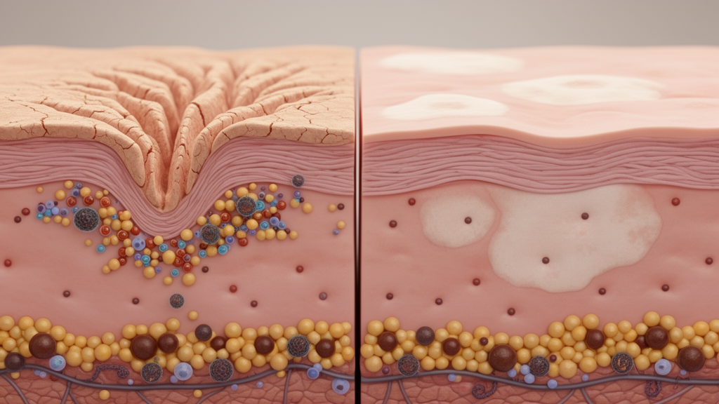
Dermatologists at Nagasaki University in Japan have documented an unusually instructive case in which two of the most recognizable immune-mediated skin diseases, psoriasis and vitiligo, occupied the very same patches of skin on a single patient. The report, published as a letter in the Journal of Cutaneous Immunology and Allergy, dissects the histopathology and immune-cell landscape of the overlapping lesions and offers a rare window into how two fundamentally different autoimmune processes can coexist, and sometimes collide, within millimeters of one another. Because psoriasis and vitiligo rarely share the same anatomical territory, the case allowed the researchers to compare inflamed and depigmented skin side by side in the same body, at the same time, under the same systemic immune conditions.
The patient was a 47-year-old man whose psoriasis vulgaris had been diagnosed 15 years earlier. After topical treatment of his psoriatic plaques, something striking happened: depigmented patches, the hallmark of vitiligo, emerged at sites that overlapped with the psoriatic lesions and then expanded in step with his overall psoriasis disease activity. When he first presented to the Nagasaki team eight years ago, he received a dual diagnosis of psoriasis and vitiligo. Oral cyclosporine, an immunosuppressant that dampens T-cell activation, was tried at a dose of 2.5 mg/kg/day, later tapered to 1 mg/kg/day, but it failed to control the psoriasis and, notably, the vitiligo continued to spread. At that point his psoriasis was severe, with a Psoriasis Area and Severity Index score of 23.2 and 20 percent of his body surface area affected, while he showed no joint symptoms suggestive of psoriatic arthritis.
With conventional immunosuppression failing, the clinicians turned to a more targeted weapon: risankizumab, an antibody that blocks the p19 subunit of interleukin-23, a cytokine central to the psoriatic inflammatory cascade. The patient received 150 mg subcutaneously at baseline, again one month later, and then every three months. The response was dramatic on the psoriatic side of the ledger. Within four months of treatment, his psoriasis improved markedly, and over five years of continuous therapy he experienced no new psoriatic lesions. The vitiligo, however, told a different story. It persisted and even expanded mildly despite the sustained IL-23 blockade, a dissociation that carries important biological and therapeutic implications.
Before starting the antibody therapy, the team obtained skin biopsies from two carefully chosen sites: one lesion containing both psoriasis and vitiligo, which the authors designate PVL, and a purely vitiliginous lesion without psoriatic features, designated VL. Under the microscope, the difference was unmistakable. The co-localized lesion displayed the textbook architecture of psoriasis: parakeratosis, the retention of nuclei in the stratum corneum; elongated rete ridges plunging deep into the dermis; Munro’s microabscesses, clusters of neutrophils trapped within the horny layer; and a dense inflammatory infiltrate in the dermis. The pure vitiligo lesion, by contrast, showed only mild epidermal hyperplasia with a limited superficial dermal infiltrate. Critically, neither lesion contained any detectable melanin or melanocytes when stained with Fontana-Masson and Melan-A, confirming complete melanocyte loss in both territories.
The immunological deep-dive focused on a panel of inflammatory cell markers: CD3 for total T cells, CD4 for helper T cells, CD8 for cytotoxic T cells, CD123 for plasmacytoid dendritic cells, and Foxp3 for regulatory T cells. In the co-localized lesion, the dermis was packed with CD3-positive and CD4-positive cells, while CD8-positive cells appeared in both the dermis and, more sparsely, in the epidermis. CD123-positive cells were distributed throughout the dermis, and Foxp3-positive regulatory T cells were present in small numbers. The pure vitiligo lesion showed only scant numbers of all five cell types in both epidermis and dermis, a picture of relative immunological quiet.
Quantitative analysis sharpened the contrast. Counting positively stained cells in three representative microscopic fields, the researchers found roughly eight times more CD3-positive T cells in the co-localized lesion than in the pure vitiligo lesion, 335 plus or minus 211 versus 42 plus or minus 36 cells per 100-times field. Plasmacytoid dendritic cells, identified by CD123, were nearly six times more abundant in the psoriasis-containing lesion, at 32 plus or minus 19.2 versus 5.6 plus or minus 4.6 cells per field. Interestingly, when the authors calculated the proportion of regulatory T cells as the Foxp3-to-CD3 ratio, the two lesions looked broadly similar: 45.4 percent in the co-localized lesion versus 37.0 percent in the pure vitiligo lesion. The message, the authors argue, is that the co-localized site simply hosted far more inflammation, while the overall cellular composition remained comparable between the two, likely reflecting the intrinsic inflammatory nature of psoriasis rather than a disease-specific interaction between the two conditions.
Several threads of prior research help make sense of these numbers. A earlier report by Ono and colleagues examined coexisting vitiligo and psoriasis lesions and found that the proportion of regulatory T cells among CD3-positive T cells increased from vitiliginous to psoriatic tissue, a pattern consistent with the Nagasaki findings. The abundance of CD123-positive cells in the co-localized lesion also points to plasmacytoid dendritic cells as players in psoriatic inflammation, echoing a landmark study showing that these cells can initiate psoriasis through production of interferon-alpha. But here lies the biological irony: plasmacytoid dendritic cells and type I interferons have also been implicated in driving progressive vitiligo. The same innate immune pathway that helps ignite psoriasis may simultaneously fuel the destruction of melanocytes, potentially explaining how the two diseases can coexist in one patch of skin and why blocking IL-23 alone left the depigmentation untouched.
The clinical course raises the question of causality. Psoriasis preceded the vitiligo in this patient, and the absence of melanocytes within the inflamed areas supports the idea that chronic psoriatic inflammation may have inflicted secondary damage on melanocytes. Yet the picture is not that simple. The psoriatic changes were concentrated mainly in the central regions of the vitiligo patches rather than at their actively expanding edges, which does not fully fit a straightforward Köbner phenomenon, the process by which trauma or inflammation triggers psoriatic lesions in previously unaffected skin. The authors also note a sobering precedent: a separate case report described de novo vitiligo emerging during IL-23p19 inhibitor therapy, suggesting that this drug class may not suppress vitiligo-associated inflammation and may, in some patients, even contribute to its development.
The authors are candid about the limits of their evidence. This is a single case report, with biopsy sampling restricted to two sites and without detailed functional immunological analyses such as cytokine profiling or single-cell sequencing. The findings neither confirm nor exclude a Köbner phenomenon as the mechanism linking the two diseases. Still, the case carries practical weight for clinicians. It demonstrates that a highly effective IL-23 pathway blockade can clear psoriasis while leaving vitiligo to pursue its own course, reinforcing the concept that these diseases, though both immune-mediated, run on partly separable molecular circuits. It also adds to a small but growing literature on co-localized psoriasis and vitiligo, a phenomenon rare enough that each well-documented case meaningfully expands the field’s understanding.
What makes the report resonate beyond dermatology is the broader immunological lesson it encodes. The skin can simultaneously host a Th17-driven, IL-23-dependent inflammatory program and a cytotoxic, interferon-influenced attack on pigment cells, with regulatory T cells present in similar proportions in both settings. Dissecting which signals are shared and which are disease-specific, particularly the ambiguous role of plasmacytoid dendritic cells and type I interferons, may inform future therapies aimed at calming one autoimmune process without unleashing another. For now, the Nagasaki case stands as a vivid reminder that the immune system’s battles in the skin are not always mutually exclusive, and that the same patch of skin can be a battlefield for two wars at once.
Subject of Research: Co-localized psoriasis and vitiligo in a single patient, with immunohistochemical analysis of immune cell infiltration and the differential response to IL-23 blockade
Article Title: A coexistence case of psoriasis and vitiligo: insights into possible pathomechanisms
Article References: A coexistence case of psoriasis and vitiligo: insights into possible pathomechanisms. (n.d.). https://doi.org/10.3389/jcia.2026.17495
Image Credits: AI Generated
Keywords: psoriasis, vitiligo, IL-23, risankizumab, plasmacytoid dendritic cells, Köbner phenomenon, regulatory T cells, interferon, melanocytes, immunohistochemistry, case report, dermatology
News Source: Ophelia Keating. (October 16, 2026). When Psoriasis and Vitiligo Collide: A Rare Case Reveals Clashing Immune Pathways in Human Skin. Scienmag.