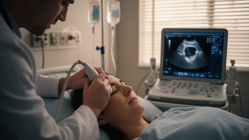
When a stroke floods the brain with damage, one of the deadliest threats is invisible: rising pressure inside the skull. Elevated intracranial pressure can crush delicate brain tissue, shift it across rigid internal membranes, and cut off blood supply to regions that were still salvageable hours earlier. Detecting this danger early usually requires drilling into the skull to place an invasive pressure monitor, a procedure that carries risks of bleeding and infection. Now, a prospective study from Cairo suggests that a simple ultrasound probe pressed against the closed eyelid may identify dangerously high intracranial pressure in patients with ischemic stroke or hypertensive intracerebral hemorrhage, and may even flag who is likely to survive their hospital stay.
The research, published in the journal Neurocritical Care, was conducted by a team of critical care physicians, neurologists, radiologists, and clinical pharmacists affiliated with Kasr Al-Ainy Medical School at Cairo University Hospital and collaborating institutions in Egypt. The investigators enrolled 100 neurocritical intensive care unit patients with acute, nontraumatic brain injury, either ischemic stroke caused by a blocked vessel or intracerebral hemorrhage driven by hypertension. All patients had severely depressed consciousness, scoring below 9 on the Glasgow Coma Scale, the standard measure of awareness that ranges from 3 for unresponsive to 15 for fully alert. Each patient underwent a computed tomography scan of the brain on admission and a bedside ultrasound measurement of the optic nerve sheath diameter.
The logic behind the technique rests on a quirk of anatomy. The optic nerve is not a free-floating cable; it is wrapped in a sleeve of dura mater, the same tough membrane that encases the brain, and this sleeve communicates with the space around the brain where cerebrospinal fluid circulates. When intracranial pressure rises, fluid is pushed along the nerve, and the sheath distends like a pressurized hose. Because the sheath is most distensible just behind the eyeball, a few millimeters of measurement through the orbit can serve as a proxy for pressure deep inside the skull. The probe emits high-frequency sound waves, receives the echoes, and displays the nerve and its sheath in cross-section, allowing clinicians to measure the diameter in seconds at the bedside.
To establish what a normal reading looks like, the researchers also recruited 60 age- and sex-matched healthy volunteers. Importantly, these controls were included only to provide descriptive reference values for physiological optic nerve sheath diameter; they were not part of the primary diagnostic or prognostic analyses. The results formed a striking staircase. Healthy volunteers averaged 4.62 millimeters, with a standard deviation of 0.33. Stroke patients whose CT scans showed no radiologic signs of elevated pressure averaged 5.52 millimeters. Patients whose scans did show CT-based surrogates of raised intracranial pressure averaged 6.50 millimeters, nearly two millimeters wider than the healthy cohort.
Because the study used CT-based radiologic surrogates as the reference standard rather than invasive pressure monitors, the team could calculate how well the ultrasound measurement discriminated between patients with and without imaging evidence of elevated pressure. Using a cutoff derived from their own cohort of 6.00 millimeters or greater, the measurement achieved 86.7 percent sensitivity, meaning it correctly identified nearly nine out of ten patients with CT-positive elevated pressure, and 85.0 percent specificity, correctly clearing about nine out of ten patients without it. The positive predictive value was 89.7 percent, the negative predictive value 81.0 percent, and overall accuracy 86.0 percent. The area under the receiver operating characteristic curve, a summary of discrimination that ranges from 0.5, no better than chance, to 1.0, perfect, reached 0.901, a figure that places the technique in the range of genuinely useful clinical tests.
Diagnostic accuracy alone would be noteworthy, but the prognostic findings may carry even greater weight. In an exploratory survival analysis, patients whose optic nerve sheath diameter exceeded the 6.00 millimeter threshold had significantly higher in-hospital mortality than those below it, with a log-rank p-value of 4.77 multiplied by 10 to the power of minus 13, an extraordinarily small probability of observing such a survival difference by chance. In other words, a single number obtained with a handheld probe in the first hours of admission carried information about who would live or die during the hospitalization.
The researchers did not stop at raw associations. Elevated intracranial pressure tends to accompany other adverse features, including deeper coma and lower blood pressure, so a crude link between sheath diameter and outcome could simply reflect that patients with big hemorrhages have both wide sheaths and poor prognoses. To address this, the team ran exploratory multivariable models adjusting for age, body mass index, Glasgow Coma Scale score, and systolic blood pressure. Even after these adjustments, an optic nerve sheath diameter above the threshold remained independently associated with CT-positive elevated intracranial pressure, supporting the interpretation that the ultrasound signal reflects pressure itself rather than merely the severity of the underlying brain insult.
Why does this matter globally? Invasive intracranial pressure monitoring requires neurosurgical expertise, sterile operating conditions, and equipment that many hospitals in low- and middle-income settings simply cannot guarantee around the clock. Egypt, like much of the world, faces a substantial burden of hypertensive intracerebral hemorrhage, a condition in which pressure dynamics evolve rapidly and every hour of unrecognized elevation worsens outcomes. A noninvasive tool that any trained clinician can deploy with a portable ultrasound machine changes the calculus of triage: it can identify patients who need aggressive pressure-lowering therapy, transfer to a facility capable of invasive monitoring, or repeated serial examinations to track an evolving crisis. The authors specifically highlight its potential for early triage and longitudinal bedside surveillance, particularly when invasive monitoring is unavailable or delayed.
The technique is not without caveats. The reference standard here was radiologic rather than a directly measured intracranial pressure in millimeters of mercury, which means the study validates ONSD against imaging surrogates rather than against the gold standard itself. The cutoff of 6.00 millimeters was derived from this specific cohort, and different populations or different ultrasound protocols may warrant different thresholds; indeed, systematic reviews and meta-analyses across traumatic brain injury, pediatric populations, and other neurological conditions have reported a spread of proposed cutoffs, reflecting variation in technique, anatomy, and patient mix. Measurement of the sheath also demands careful technique and operator training, which is why international consensus groups have published quality criteria for image acquisition and measurement. Furthermore, the survival and adjustment analyses are described by the authors as exploratory, and the prognostic signal, however striking, would need confirmation in independent cohorts before sheath diameter becomes a standard element of stroke prognostication.
Nevertheless, the study adds an important piece of evidence in a field long dominated by data from traumatic brain injury. Nontraumatic brain injury patients, those with spontaneous strokes and hemorrhages, have been underrepresented in the ultrasound literature, and this prospective cohort of 100 severely affected patients provides some of the clearest data yet that the eye can serve as a window on the injured brain. The stepwise increase from healthy controls to CT-negative patients to CT-positive patients is internally consistent, the discrimination statistics are strong, and the mortality signal is dramatic. If larger, multicenter studies reproduce these findings, a probe, a dollop of gel, and a closed eyelid could become part of the standard first assessment of every comatose stroke patient, turning a routine ultrasound exam into a rapid, radiation-free estimate of one of neurocritical care’s most feared variables.
Subject of Research: Optic nerve sheath diameter ultrasonography for detecting elevated intracranial pressure and prognosis in ischemic stroke and hypertensive intracerebral hemorrhage
Article Title: Diagnostic and Prognostic Value of Sonographic Optic Nerve Sheath Diameter in Patients with Ischemic Stroke or Hypertensive Intracerebral Hemorrhage
Article References: Diagnostic and Prognostic Value of Sonographic Optic Nerve Sheath Diameter in Patients with Ischemic Stroke or Hypertensive Intracerebral Hemorrhage. (n.d.). https://doi.org/10.1007/s12028-026-02697-8
Image Credits: AI Generated
DOI: 10.1007/s12028-026-02697-8
Keywords: optic nerve sheath diameter, intracranial pressure, ultrasonography, ischemic stroke, intracerebral hemorrhage, neurocritical care, diagnostic accuracy, prognosis, point-of-care ultrasound, Glasgow Coma Scale, ICU monitoring, computed tomography
News Source: Cassandra Pierce. (October 16, 2026). Bedside Eye Ultrasound Reveals Dangerous Brain Pressure in Stroke Patients. Scienmag.