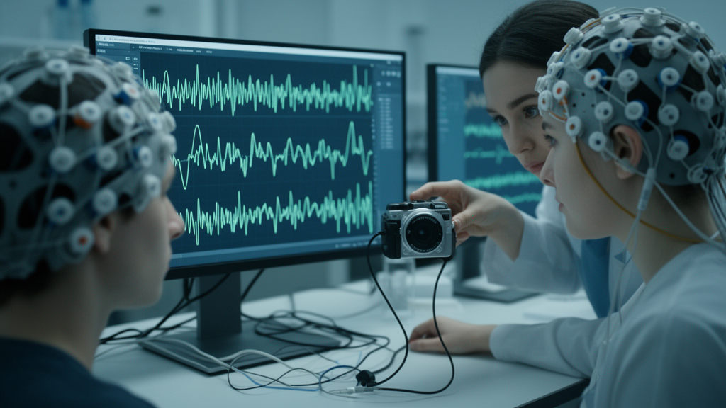
Deep learning has swept through brain-computer interface research with the promise of superhuman pattern recognition, and nowhere is that enthusiasm more visible than in motor imagery BCIs, systems that read a user’s imagined hand movements directly from scalp electrodes. But a new study from researchers at Inria and the University of Bordeaux delivers a sobering warning: some of the impressive accuracy gains reported for neural networks decoding electroencephalography, or EEG, may come not from reading thoughts at all, but from the networks quietly latching onto the visual flashes that tell participants what to imagine. In experiments published in Frontiers in Neuroergonomics, the team showed that convolutional and Transformer-based EEG models scored substantially higher when the brief visual cue that instructs the user was left inside the analyzed signal window, while a classic machine learning pipeline remained essentially indifferent to its presence.
The investigation focused on one of the most widely studied BCI paradigms, in which users imagine moving their left or right hand and the system must decide which. The genuine neural signature of such imagery is a modulation of sensorimotor rhythms over the motor cortex, specifically event-related desynchronization and synchronization in the mu and beta frequency bands, roughly 8 to 30 hertz, observed contralaterally at electrodes C3 and C4. Yet every trial in the standard laboratory protocol begins with a visual cue, typically an arrow pointing left or right, and the brain’s response to that arrow, a visual evoked potential, is itself a strong, time-locked electrical event. The researchers asked a deceptively simple question: when the analysis window includes the cue onset, are classifiers decoding the imagery, or are they decoding the arrow?
To answer it, the team evaluated three very different classification pipelines on two large open-access EEG datasets, Dreyer2023 with 85 participants and Lee2019 with 54. The first was CSP combined with Linear Discriminant Analysis, the workhorse of traditional BCI research, which spatially filters the signal and then classifies the averaged band power, discarding temporal structure. The second was DeepConvNet, a convolutional neural network with roughly 283,000 parameters that learns both temporal and spatial filters directly from raw data. The third was REVE, a Transformer-based EEG foundation model pretrained on more than 6,000 hours of recordings from over 25,000 subjects and containing about 69 million parameters. Each model was trained twice per dataset under two epoching conditions: windows that either started at the visual cue onset or began half a second later, deliberately excluding it, with all other preprocessing held constant.
The headline result was striking. For DeepConvNet, including the cue onset raised cross-subject classification accuracy by 12.6 percent on Dreyer2023 and 5.4 percent on Lee2019; for REVE, the corresponding gains were 8.9 and 5.3 percent. All of these differences were highly significant under paired t-tests corrected for multiple comparisons. On its face, that would look like free performance, exactly the kind of boost that might tempt a benchmark winner. But the authors then turned to explainable artificial intelligence tools, applying saliency maps, Integrated Gradients, and DeepLIFT to visualize which channels and time points drove each model’s decisions, and the story changed dramatically.
Attribution analysis revealed that the deep networks trained with the cue onset concentrated their attention almost entirely on the first half second after stimulus presentation, and on parietal electrodes such as P3 and P4, regions associated with visual processing rather than motor control. In the Dreyer2023 data, P3 and P4 alone accounted for roughly 18.2 and 12.0 percent of the model’s decision, and the sharpest temporal attribution peak appeared around 190 milliseconds after cue onset, precisely where visual evoked potentials are expected. When the cue was excluded, the picture inverted: attribution spread across the whole trial, focused on C3 and C4, and resembled the classic contralateral ERD patterns described in the neuroscience literature. The networks were not reading imagined movements when the cue was present; they were reading the visual response to the cue.
The classical CSP+LDA pipeline behaved completely differently. In the within-subject setting, mean accuracies were nearly identical with and without the cue, 67.8 versus 67.9 percent on Dreyer2023, with no significant difference, and the extracted CSP patterns stayed anchored over sensorimotor cortex in both conditions. In the cross-subject setting, CSP+LDA actually performed slightly better when the cue was removed. The authors attribute this asymmetry to the mathematics of the two approaches. CSP averages signal power over the entire epoch, cannot exploit slow event-related potentials, and was fed signals band-pass filtered to 8 to 30 hertz, which strips out most cue-related low-frequency components. Deep learning models, by contrast, process broadband signals from 0.5 to 40 hertz and can learn arbitrary temporal filters, making them exquisitely sensitive to transient visual evoked potentials that the traditional method simply cannot see.
Independent ERP analyses confirmed the mechanism. Time-locked averages revealed clear visual evoked potentials around 100 to 200 milliseconds after cue onset, plus a central-parietal positivity around 300 to 500 milliseconds consistent with a P300 component, and a class-discriminant early VEP was most pronounced in the subjects whose accuracy changed most between conditions. Notably, the researchers also tried a simpler fix: deleting the parietal channels most likely to capture visual responses. It failed. Even without those electrodes, DeepConvNet still gained 9.2 percent from including the cue, almost certainly because volume conduction spreads visual activity across the scalp.
The practical implications reach far beyond one experimental design choice. In real-world, asynchronous BCI use, there is no visual cue, because users initiate commands themselves, so a classifier built on cue-related potentials would simply fail outside the laboratory. Even in cue-guided applications such as BCI-based stroke rehabilitation, rewarding attention to arrows rather than genuine sensorimotor modulation could actively undermine therapy. The problem is not confined to this study; the authors point out that prominent benchmarks, including the NeurIPS 2021 BEETL competition, have provided tutorial code that epochs data from the cue onset, potentially biasing reported results across the field.
The team’s recommendations are concrete: exclude visual cue periods from EEG input windows for deep learning models, run ERP analyses to determine cue-locked latencies before choosing windows, and above all make explainability tools such as attribution maps a routine part of BCI model evaluation rather than an optional extra. Higher accuracy, they conclude, does not guarantee neurophysiological validity. In a field racing to deploy neural networks on fragile, noisy brain signals, that message may be the most important finding of all: before trusting a leaderboard, ask what the model is actually looking at.
Subject of Research: Visual cue-induced bias in deep learning versus standard machine learning EEG classifiers for motor imagery brain-computer interfaces
Article Title: Visual cues in MI-BCI induce biases in EEG classification models with deep learning but not with standard machine learning
Article References: Trocellier, D., Kojima, S., N'kaoua, B., & Lotte, F. (2026). Visual cues in MI-BCI induce biases in EEG classification models with deep learning but not with standard machine learning. Frontiers in Neuroergonomics, 7, Article 1804143. https://doi.org/10.3389/fnrgo.2026.1804143
Image Credits: AI Generated
DOI: 10.3389/fnrgo.2026.1804143
Keywords: brain-computer interface, EEG, deep learning, motor imagery, visual evoked potentials, CSP, LDA, DeepConvNet, explainable AI, classification bias, neurophysiology, machine learning
News Source: Blake Davidson. (October 15, 2026). Visual Cues Quietly Inflate Deep Learning Accuracy in Brain-Computer Interfaces, Study Finds. Scienmag.