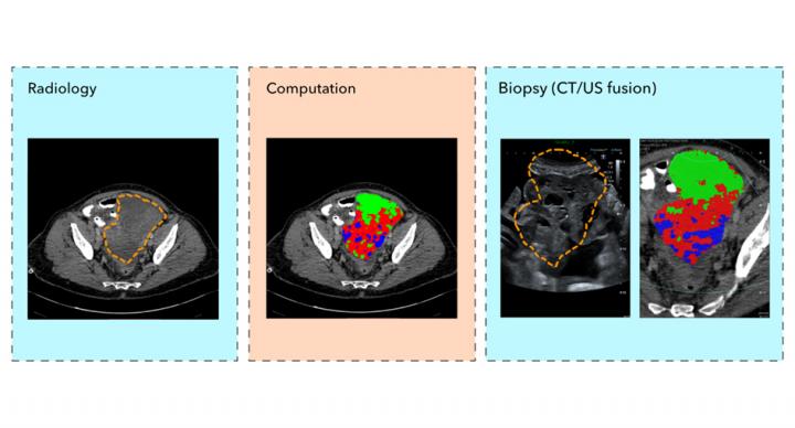
Credit: Evis Sala/University of Cambridge
A new advanced computing technique using routine medical scans to enable doctors to take fewer, more accurate tumour biopsies, has been developed by cancer researchers at the University of Cambridge.
This is an important step towards precision tissue sampling for cancer patients to help select the best treatment. In future the technique could even replace clinical biopsies with ‘virtual biopsies’, sparing patients invasive procedures.
The research published in European Radiology shows that combining computed tomography (CT) scans with ultrasound images creates a visual guide for doctors to ensure they sample the full complexity of a tumour with fewer targeted biopsies.
Capturing the patchwork of different types of cancer cell within a tumour – known as tumour heterogeneity – is critical for selecting the best treatment because genetically-different cells may respond differently to treatment.
Most cancer patients undergo one or several biopsies to confirm diagnosis and plan their treatment. But because this is an invasive clinical procedure, there is an urgent need to reduce the number of biopsies taken and to make sure biopsies accurately sample the genetically-different cells in the tumour, particularly for ovarian cancer patients.
High grade serous ovarian (HGSO) cancer, the most common type of ovarian cancer, is referred to as a ‘silent killer’ because early symptoms can be difficult to pick up. By the time the cancer is diagnosed, it is often at an advanced stage, and survival rates have not changed much over the last 20 years.
But late diagnosis isn’t the only problem. HGSO tumours tend to have a high level of tumour heterogeneity and patients with more genetically-different patches of cancer cells tend to have a poorer response to treatment.
Professor Evis Sala from the Department of Radiology, co-lead CRUK Cambridge Centre Advanced Cancer Imaging Programme, leads a multi-disciplinary team of radiologists, physicists, oncologists and computational scientists using innovative computing techniques to reveal tumour heterogeneity from standard medical images. This new study, led by Professor Sala, involved a small group of patients with advanced ovarian cancer who were due to have ultrasound-guided biopsies prior to starting chemotherapy.
For the study, the patients first had a standard-of-care CT scan. A CT scanner uses x-rays and computing to create a 3D image of the tumour from multiple image ‘slices’ through the body.
The researchers then used a process called radiomics – using high-powered computing methods to analyse and extract additional information from the data-rich images created by the CT scanner – to identify and map distinct areas and features of the tumour. The tumour map was then superimposed on the ultrasound image of the tumour and the combined image used to guide the biopsy procedure.
By taking targeted biopsies using this method, the research team reported that the diversity of cancer cells within the tumour was successfully captured.
Co-first author Dr Lucian Beer, from the Department of Radiology and CRUK Cambridge Centre Ovarian Cancer Programme, said of the results: “Our study is a step forward to non-invasively unravel tumour heterogeneity by using standard-of-care CT-based radiomic tumour habitats for ultrasound-guided targeted biopsies.”
Co-first author Paula Martin-Gonzalez, from the Cancer Research UK Cambridge Institute and CRUK Cambridge Centre Ovarian Cancer Programme, added: “We will now be applying this method in a larger clinical study.”
Professor Sala said: “This study provides an important milestone towards precision tissue sampling. We are truly pushing the boundaries in translating cutting edge research to routine clinical care.”
Fiona Barve (56) is a science teacher who lives near Cambridge. She was diagnosed with ovarian cancer in 2017 after visiting her doctor with abdominal pain. She was diagnosed with stage 4 ovarian cancer and immediately underwent surgery and a course of chemotherapy. Since March 2019 she has been cancer free and is now back to teaching three days a week.
“I was diagnosed at a late stage and I was fortunate my surgery, which I received within four weeks of being diagnosed, and chemotherapy worked for me. I feel lucky to be around,” said Barve.
“When you are first undergoing the diagnosis of cancer, you feel as if you are on a conveyor belt, every part of the journey being extremely stressful. This new enhanced technique will reduce the need for several procedures and allow patients more time to adjust to their circumstances. It will enable more accurate diagnosis with less invasion of the body and mind. This can only be seen as positive progress.”
###
This feasibility study, involving researchers from the Department of Radiology, CRUK Cambridge Institute, Addenbrooke’s Hospital, Cambridge University Hospitals NHS Foundation Trust, and collaborators at Cannon, was facilitated through the CRUK Cambridge Centre Integrated Cancer Medicine programme.
The goal of Integrated Cancer Medicine is to revolutionise cancer treatment using complex data integration. Combining and integrating patient data from multiple sources – blood tests, biopsies, medical imaging, and genetic tests – can inform and predict the best treatment decisions for each individual patient.
The study was funded by Cancer Research UK and The Mark Foundation for Cancer Research.
Reference
Lucian Beer, Paula Martin-Gonzalez et al. Ultrasound-guided targeted biopsies of distinct CT based radiomic tumour habitats: proof of concept. European Radiology; 14 Dec 2020; DOI: 10.1007/s00330-020-07560-8
Media Contact
Craig Brierley
[email protected]
Original Source
https:/
Related Journal Article
http://dx.


