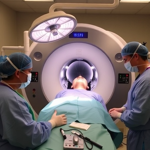
Surgeons at Vanderbilt University Medical Center’s Department of Otolaryngology-Head and Neck Surgery have pioneered a groundbreaking surgical procedure in the United States utilizing the most advanced generation of intraoperative positron emission tomography (PET) combined with computed tomography (CT) scanning technology. This innovation aims to dramatically improve surgical precision and ultimately enhance patient outcomes, specifically in surgeries involving head and neck cancers. The technique employs the Aura 10 scanner—a state-of-the-art hybrid imaging device developed by Belgian surgical technology company Xeos—which integrates real-time functional and anatomical imaging directly into the operating room.
Dr. Michael Topf, an associate professor specializing in Otolaryngology-Head and Neck Surgery at Vanderbilt, spearheaded the initial surgery employing this novel imaging approach. He and his team have since performed multiple surgeries as part of an extensive clinical study involving up to 50 patients. The study’s principal objective is to evaluate the long-term feasibility and clinical benefits of incorporating intraoperative PET-CT to assess tumor resections during surgery, aiming to set a new standard in cancer care.
Intraoperative PET-CT represents a significant technological leap by enabling surgeons to conduct detailed margin analysis in real-time. Traditionally, resected tumor specimens are sent to pathology labs, where comprehensive microscopic evaluations can take days to finalize before confirming whether the surgical margins are clean of cancer cells. By contrast, this new method transports the resected tissue immediately to the nearby scanner within the operating suite, supplying surgeons with rapid and critical feedback on the completeness of the cancer removal.
The innovative imaging modality capitalizes on the strengths of PET and CT technologies in unison. PET provides vital functional insights by detecting metabolic activity, using radiotracers that highlight malignant tissues due to their elevated glucose uptake. Meanwhile, the CT scan offers detailed anatomical views, outlining the tumor and its surroundings with high spatial resolution. This fusion delivers a comprehensive image, enabling surgeons to identify residual malignant cells that could remain undetectable by visual inspection alone.
During the surgical procedure, patients receive an injection of fluorodeoxyglucose (FDG), a radiopharmaceutical that mimics glucose. Cancerous cells, being metabolically hyperactive, absorb and retain FDG more than normal tissues, causing the tumor to “light up” on the PET scan. Once the tumor is excised, the specimen is scanned immediately with the Aura 10 device, and the imaging data are analyzed to determine if the edges of the removed tissue—termed margins—are free from cancerous cells. This immediate feedback empowers surgeons to decide whether additional tissue needs excision to ensure a negative margin and reduce the risk of recurrence.
Dr. Topf emphasized that the ongoing research not only tests the feasibility of using intraoperative PET-CT in the operating room but also rigorously compares its accuracy against the pathology lab’s gold standard of microscopic tissue analysis. Validation of this technology could revolutionize head and neck oncology surgeries by providing surgeons greater confidence during resections, potentially improving cure rates and sparing patients from the morbidity of incomplete removals or excessive surgery.
Nicole Jones, the research coordinator IV in the otolaryngology laboratory, highlighted the transformative clinical implications of incorporating this technology. She noted that current practices involve a time lag between surgery and pathology results, often leading to delayed decisions about follow-up treatments or surgeries. The immediate intraoperative imaging poster presented by the Aura 10 scanner could profoundly impact patient care by drastically shortening this feedback loop, allowing surgical teams to tidy up any residual cancer in a single operative session.
Such innovation redefines surgical workflows by keeping everything within the operative environment. Surgeons no longer need to leave the sterile operating theater to consult pathology results; instead, the entire cycle of tumor removal and assessment occurs seamlessly and in real-time. This integrated model is poised to enhance surgical precision, reduce reoperation rates, and improve overall patient outcomes. It also embodies a bold leap towards more personalized, data-driven surgical oncology.
From the patient perspective, Dr. Topf noted that participation in this clinical research offers a unique and compelling opportunity to benefit from cutting-edge technology without additional hospital visits or invasive testing. Using intraoperative PET-CT represents a convergence of advanced imaging and surgical techniques designed to elevate the standard of care and instill confidence in both surgeons and patients that cancer removal is as thorough and minimally invasive as possible.
The implications of this technology extend beyond head and neck cancer surgeries, potentially applying to numerous other oncologic and surgical disciplines where accurate margin assessment is critical. The real-time, intraoperative visualization of cancer metabolism and localization offers a window into personalized surgery, where every decision is informed by precise biological and anatomical data.
In summary, the integration of the Aura 10 intraoperative PET-CT scanner into head and neck cancer surgeries represents a paradigm shift in oncologic surgery. This novel approach promises to reduce positive surgical margins, lower recurrence rates, and accelerate clinical decision-making. The ongoing study at Vanderbilt University Medical Center, led by Dr. Michael Topf and his team, continues to evaluate the technology’s long-term viability and hopes to pave the way for widespread clinical adoption of intraoperative molecular imaging.
As the landscape of cancer surgery evolves, innovations like this that blend real-time molecular imaging with surgical practice could herald a new era in precision oncology, improving patient survival and quality of life through enhanced surgical accuracy and tailored treatment approaches. The success of this research may ultimately redefine how surgeons approach cancer resections and set new benchmarks for operative excellence worldwide.
Subject of Research: Advanced intraoperative imaging technology in head and neck cancer surgery
Article Title: Breakthrough Use of Intraoperative PET-CT in Head and Neck Cancer Surgery at Vanderbilt University
News Publication Date: (Information not provided)
Web References: (Information not provided)
References: (Information not provided)
Image Credits: Photo by Erin O. Smith, Vanderbilt University Medical Center
Keywords: Head and neck cancer, Otolaryngology, Intraoperative PET-CT, Surgical oncology, Cancer margin assessment, Real-time molecular imaging
Tags: advanced intraoperative PET-CT scan technologyAura 10 hybrid imaging deviceclinical study on tumor resectionsDr. Michael Topf otolaryngologyhead and neck cancer surgeryinnovative cancer care techniquesintraoperative imaging advancementspatient outcomes improvementreal-time imaging in surgerysurgical precision enhancementsurgical technology breakthroughsVanderbilt University Medical Center


