In the rapidly evolving landscape of cancer research, a groundbreaking study from the Max Delbrück Center for Molecular Medicine (MDC) is redefining how we understand the tumor microenvironment. Led by Professor Nikolaus Rajewsky and his team at the Berlin Institute for Medical Systems Biology, this study marries cutting-edge spatial transcriptomics with advanced extracellular matrix imaging to create a comprehensive three-dimensional map of cellular ecosystems within a lung tumor. These novel insights open unprecedented avenues for personalized cancer therapies by focusing not only on cellular composition but, crucially, on spatial organization and intercellular communication.
Tumors are no longer viewed as mere clusters of malignant cells but as complex ecosystems. The intricate interplay between cancer cells, immune cells, fibroblasts, and surrounding extracellular matrix (ECM) literally shapes disease progression and treatment responses. While traditional pathology protocols offered two-dimensional snapshots often limited to histological staining, this research leverages high-resolution single-cell spatial technologies, offering a multidimensional and molecularly precise view of tumor architecture with cellular neighborhood resolution.
Central to this advancement is the application of spatial transcriptomics, a technology that profiles RNA expression with remarkable spatial context. Unlike conventional transcriptomics, which bulk-analyzes RNA from homogenized tissue samples, spatial transcriptomics preserves the positional information of transcripts at single-cell resolution. Employing the innovative CosMx platform by NanoString, Rajewsky’s team was able to detect up to 1,000 distinct RNA molecules per cell, a quantum leap from earlier methods constrained to just a few markers. This enabled detailed profiling of over 340,000 individual cells, encompassing 18 distinct cell types within a single lung tumor, highlighting the heterogeneous cellular milieu.
However, the leap from two-dimensional to three-dimensional analysis required innovative computational solutions. The team introduced STIM, a novel algorithm designed to reconstruct 3D virtual tissue blocks by aligning multiple spatial transcriptomic datasets. STIM conceptualizes spatial transcriptomic data as digital images, applying computer vision techniques to stack and align these images, culminating in a holistic 3D tissue reconstruction. This integration underscores the power of interdisciplinary collaboration, merging computational sciences with molecular biology, and was further enhanced by expertise from Dr. Stephan Preibisch at the Howard Hughes Medical Institute.
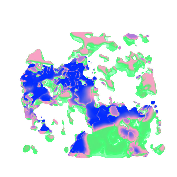
The 3D reconstructions revealed more than just cellular identities: by coupling these maps with second harmonic generation imaging, the researchers visualized key ECM components, including elastin and collagen fibers. This dual-layered imaging disclosed spatial variations in ECM composition, where elastin-rich regions correlated with healthier tissue, whereas areas rich in collagen congregated around the tumor core, signifying deleterious tissue remodeling driven by the tumor microenvironment. Such details illuminate how structural alterations to the ECM contribute to cancer progression.
Crucially, this approach illuminated dynamic cellular interactions within the tumor. Fibroblasts, cells responsible for synthesizing connective tissue, were observed in activated states remodeling the ECM, creating a scaffold supporting tumor growth. Beyond static snapshots, the data unveiled functional phenotypes and intercellular signaling, particularly mechanisms by which tumor cells suppress infiltration of immune cells. The findings emphasized the presence of immune niches surrounding the tumor core that, despite their proximity, were functionally impaired due to tumor-induced immunosuppression.
Understanding the precise molecular crosstalk that underpins immune evasion is vital. The study exposes how tumors can inhibit immune cell penetration through known checkpoint pathways, validating immunotherapy strategies that employ immune checkpoint inhibitors. By reversing this localized immune suppression, such therapies could unleash resident immune cells that are otherwise incapacitated, offering a personalized treatment strategy that conventional chemotherapy alone could not provide.
What sets this research apart is its applicability to routine clinical samples. Despite utilizing sophisticated molecular techniques, the team demonstrated that archived formalin-fixed, paraffin-embedded (FFPE) tissue sections—commonly preserved in clinical pathology labs—are amenable to this high-resolution analysis. This “Pathology 2.0” approach transcends traditional microscopy, enriching pathological examination with molecular and spatial depth, and has the potential to transform diagnostic and therapeutic decision-making in oncology.
The translational promise of this integrated spatial approach is monumental. By comprehensively mapping cellular neighborhoods and molecular signals within tumors, physicians could tailor immunotherapies and other interventions with unprecedented precision. Moreover, expanding these techniques to larger patient cohorts is underway, with ongoing analyses involving hundreds of additional samples. Such scaling will enable validation of molecular targets and foster development of broadly applicable personalized medicine protocols.
Another frontier being explored involves integrating proteomic data into the comprehensive tissue map. Collaborations with Dr. Fabian Coscia’s Spatial Proteomics Lab at MDC aim to incorporate protein activity measurements alongside RNA expression and ECM imaging. This multi-omic synergy will deepen insights into functional tumor biology, elucidate post-transcriptional regulation, and refine therapeutic target identification.
This study represents a paradigm shift in cancer research and diagnostics. By synergizing single-cell resolution spatial transcriptomics with advanced ECM imaging and novel computational reconstructions, researchers can now dissect the tumor microenvironment with molecular and spatial fidelity previously unattainable. The resulting data describe not only who is present in the tumor but where, how, and why they interact – all essential information for designing personalized therapies capable of halting tumor progression and improving patient outcomes.
Professor Rajewsky encapsulates the significance succinctly: the comprehensive data from patient tumor tissues now allow computational predictions of the molecular mechanisms driving cancer phenotypes. This predictive capacity could revolutionize oncology, moving from generalized treatments toward truly individualized interventions—a vision for precision medicine now within reach.
In essence, the collaboration between biology, computational science, and advanced imaging heralds a new era where high-tech analytical tools refine routine pathology into a powerful platform for personalized cancer care. The integration of spatial, molecular, and functional data sets a foundation for next-generation diagnostics and therapeutics, promising hope for patients facing lung cancer and other malignancies. With further validation and clinical application, these innovations will likely redefine cancer management strategies over the coming decade.
—
Subject of Research: Cells
Article Title: Combining spatial transcriptomics and ECM imaging in 3D for mapping cellular interactions in the tumor microenvironment
News Publication Date: 11-Apr-2025
Web References: 10.1016/j.cels.2025.101261
Image Credits: Rajewsky lab, Max Delbrück Center
Keywords: spatial transcriptomics, 3D tumor mapping, extracellular matrix imaging, single-cell analysis, lung cancer, tumor microenvironment, immunotherapy, computational modeling, personalized medicine
Tags: advanced cancer treatment strategiescancer research breakthroughscellular ecosystems in tumorsextracellular matrix imagingimmune cell interactions in cancerintercellular communication in tumorsmultidimensional tumor architecturepersonalized cancer therapiessingle-cell RNA profilingspatial transcriptomics technologythree-dimensional tumor analysistumor microenvironment mapping
