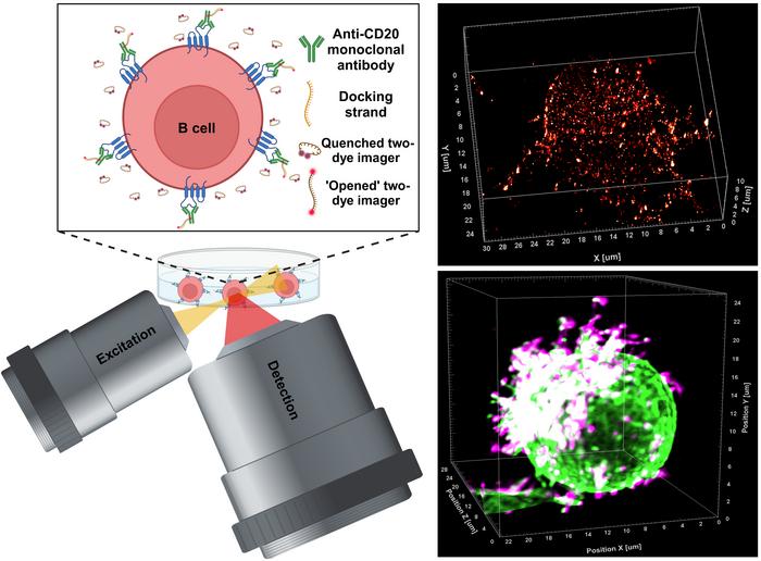

In the realm of medical research, a groundbreaking breakthrough has emerged from the laboratories of the University of Würzburg in Bavaria, Germany. The study undertaken by Professor Markus Sauer and his team signifies a major leap in the understanding and development of therapeutic antibodies. This research unveils the intricate interactions between antibodies and their target molecules, particularly in the context of B cells associated with blood cancers such as chronic lymphocytic leukaemia. The research group has developed a novel super-resolution microscopy technique known as LLS-TDI-DNA-PAINT, which allows for real-time observation of therapeutic antibodies binding to target proteins on cancer cells in three-dimensional space with unprecedented detail and accuracy.
For decades, the use of immunotherapeutic antibodies has been a significant approach in treating various tumors. One of the primary targets in such therapies is the CD20 protein present on the surface of B cells. When these antibodies bind to CD20, they induce a cascade of immune responses, ultimately leading to the destruction of malignant cells. However, despite the clinical efficacy of these antibodies, a limited understanding of their binding mechanisms and the subsequent cellular response has persisted among scientists. This gap in knowledge has now been significantly bridged by the innovative research coming out of Würzburg.
Through the application of the LLS-TDI-DNA-PAINT technique, researchers were able to visualize the dynamics of therapeutic antibodies in real time. They scrutinized how these antibodies not only adhere to CD20 molecules but also influence the structural behavior of B cells during this critical interaction. Notably, the study shared insights about how the binding of these antibodies leads to an observable phenotypic change in B cells, adopting what the researchers describe as a “hedgehog shape.” This transformation is a result of the antibodies crosslinking CD20 proteins, activating complement systems, and thus promoting targeted immune responses.
The researchers conducted these observations using the Raji B cell line, a derivative of human Burkitt’s lymphoma, recognized for its relevance in cancer studies. This particular line serves as a model for assessing the responses of B cells to therapeutic antibodies. The research team carried out experiments with four distinct therapeutic antibodies: RTX, OFA, OBZ, and 2H7, all known for their roles in targeting CD20. The results of their studies revealed that regardless of the type of antibody employed—whether classed as type I or II—the response mechanism remains strikingly similar, contradicting previous assumptions within the scientific community.
Furthermore, the binding of therapeutic antibodies was shown to occur predominantly at specialized membrane regions of the B cells, specifically microvilli. These microscopic protrusions not only act as platforms for protein interaction but also enhance the structural integrity of the B cell surface during therapeutic engagement. The observed polarization of B cells in conjunction with the stabilization of these microvilli reinforces the hypothesis that the architecture of the cell membrane plays a substantial role in the immune response. Such findings illuminate not just the mechanics of antibody binding but also hint at the potential for improving therapeutic strategies by modulating these interactions.
Another remarkable aspect of this study is the potential implication of the hedgehog-shaped B cells in fostering inter-cellular communication within the immune system. The researchers postulate that this morphological alteration might be instrumental in facilitating the formation of immunological synapses, prompting B cells to engage with macrophages and natural killer cells. This conjecture pushes the boundaries of current understanding and emphasizes the necessity for further inquiries into the roles these activated B cells play in orchestrating broader immune responses.
The establishment of the LLS-TDI-DNA-PAINT method opens pathways for an in-depth exploration of therapeutic mechanisms at a molecular level. This groundbreaking approach paves the way for future studies aimed at unraveling the specificities of cell-surface interaction and the multifaceted processes involved in immune recognition and destruction of cancerous cells. Armed with this novel visualization technology, researchers are now equipped to scrutinize even minute details of antibody binding dynamics, offering an unparalleled platform for advancing personalized immunotherapies.
As the study progresses, the researchers at the University of Würzburg intend to delve deeper into the implications their findings may have on the future of antibody-based cancer therapies. With the advancements made through the LLS-TDI-DNA-PAINT technique, there lies a promising horizon where therapeutic antibodies can be tailored and innovated based on direct observations of their interactions within the tumor microenvironment.
In summary, this significant research from Würzburg not only challenges existing paradigms within the therapeutic antibody field but also showcases the power of cutting-edge microscopy techniques in revolutionizing our understanding of cancer biology. As researchers continue their explorations, the hope for enhanced therapeutic strategies and improved patient outcomes remains high, providing a beacon of hope in the ongoing battle against blood cancers and beyond.
Subject of Research: Cells
Article Title: Decoding the molecular interplay of CD20 and therapeutic antibodies with fast volumetric nanoscopy
News Publication Date: 9-Jan-2025
Web References: Journal Article
References: N/A
Image Credits: Arindam Ghosh / University of Wuerzburg
Keywords: super-resolution microscopy, therapeutic antibodies, B cells, CD20, cancer research, immunotherapy, LLS-TDI-DNA-PAINT, chronic lymphocytic leukaemia, Burkitt’s lymphoma, immune response, molecular interactions.


