


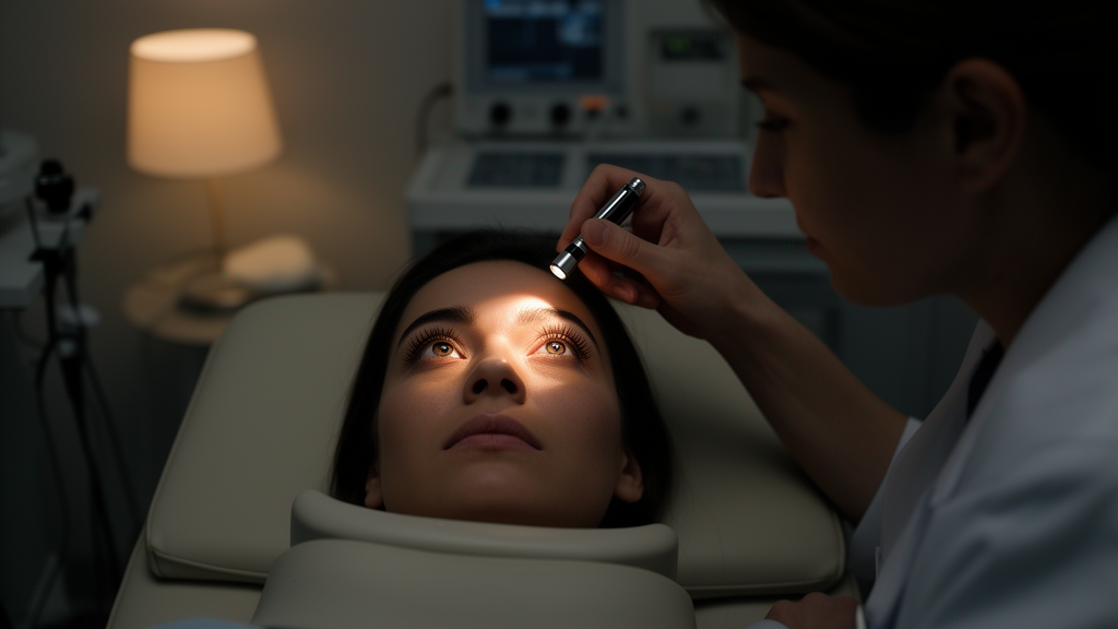
Unequal pupils in an intensive care patient are traditionally read as an alarm for intracranial hemorrhage, stroke, or transtentorial herniation. A new report argues that in patients lying face down, the finding is often far more benign. Published in Eye & ENT Research, the article draws on two case reports and a review of the literature to describe prone positioning–associated anisocoria as a diagnostic pitfall that can lead clinicians toward unnecessary emergency neuroimaging and invasive interventions.
Prone positioning itself is well established: it improves survival in severe respiratory failure, particularly acute respiratory distress syndrome. But the posture can be followed by anisocoria, and the new work shows that benign, position-related mechanisms can explain the pupillary change. In one reported case, anisocoria developed after roughly 18 hours of prone positioning and turned out to be angle-closure glaucoma. In an anatomically predisposed patient, gravity shifted the lens–iris diaphragm anteriorly, triggering pupillary block and a sharp rise in intraocular pressure. That mechanism required targeted glaucoma management rather than repositioning alone.
The second case followed a different pathway. After about eight hours prone, the patient developed miosis with ptosis—the classic signature of Horner syndrome—reflecting transient compression of the sympathetic pathway. The finding resolved completely about 45 minutes after the patient was returned to the supine position, underscoring how quickly a position-dependent compression can reverse once the mechanical trigger is removed.
From these cases, the authors propose a simplified diagnostic algorithm. Anisocoria without new neurological deficits after prone positioning is usually benign and positional, and repositioning should be the first diagnostic and therapeutic step. Immediate neuroimaging is reserved for patients with red flags such as altered consciousness, focal deficits, or loss of brainstem reflexes. Recognizing these mechanisms, the authors argue, can prevent unnecessary emergency imaging and invasive procedures in critically ill patients.
The practical message for intensive care teams is that the prone position itself generates differential diagnostic considerations that supine medicine rarely encounters. Gravitational effects on the eye and on peripheral sympathetic nerves can mimic the pupillary signs that ordinarily demand urgent brain imaging. Distinguishing a positional, self-limiting pupillary change from a true neurological emergency depends on the accompanying clinical picture—whether new deficits, altered consciousness, or brainstem reflex abnormalities are present—rather than on the anisocoria alone.
Subject of Research: Benign causes of anisocoria in prone-positioned critically ill patients
Article Title: Anisocoria after prone positioning: A benign finding not to be mistaken for brain herniation
Article References: Anisocoria after prone positioning: A benign finding not to be mistaken for brain herniation. Original publication (Bibliographic details unavailable.)
Image Credits: AI Generated
DOI: Not provided
Keywords: anisocoria, prone positioning, angle-closure glaucoma, Horner syndrome, critical care, ARDS, pupillary block, intraocular pressure, neuroimaging, diagnostic algorithm, ICU, brain herniation
News Source: Ophelia Keating. (October 29, 2026). Unequal Pupils After Prone Positioning May Signal Glaucoma or Horner Syndrome, Not Herniation. Scienmag.



