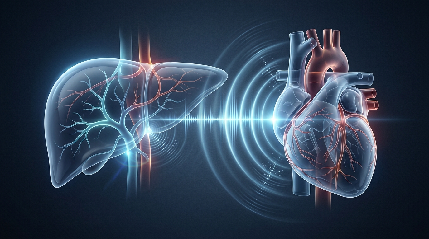
When the heart fails, the liver often pays the price. Blood backs up through the right side of the heart, pressure builds in the veins that drain the liver, and the organ slowly becomes congested, stiff, and in some cases scarred. Clinicians have long recognized this heart-liver interplay, now called cardiohepatic syndrome, yet cardiac and hepatic abnormalities are still frequently evaluated in parallel rather than as one connected hemodynamic process. A new review published in Cardiovascular Ultrasound argues that this fragmented approach is outdated and proposes a practical, sequential ultrasound pathway to evaluate the syndrome at the bedside.
The review, led by Yin-Xia Li and Rui Shen with colleagues, frames cardiohepatic syndrome as a bidirectional interaction. Heart failure is a systemic syndrome in which venous congestion and impaired perfusion can produce clinically important hepatic dysfunction. In chronic heart failure, the dominant phenotype is often congestive hepatopathy, whereas acute hemodynamic deterioration can produce hypoxic or acute cardiogenic liver injury. These phenotypes may coexist, and their severity varies over time. The relationship is not purely one-way either: hepatic inflammation, fibrosis, altered protein synthesis, and metabolic dysfunction may amplify cardiovascular risk and complicate heart failure management.
The pathophysiology follows a clear mechanical logic. Elevated right atrial pressure is transmitted through the inferior vena cava and hepatic veins to the hepatic sinusoids, the liver’s microscopic filtration channels. Persistent sinusoidal hypertension causes centrilobular congestion, hepatocyte compression, reduced oxygen diffusion, and progressive extracellular matrix remodeling. At the same time, reduced cardiac output decreases hepatic arterial perfusion and may worsen centrilobular hypoxia. The combined effect is greatest in patients with severe tricuspid regurgitation, pulmonary hypertension, right ventricular dysfunction, constrictive physiology, or advanced biventricular heart failure. Experimental models show that sustained cardiac dysfunction can induce hepatic fibrosis and that right-sided pressure overload produces congestion, necrosis, and matrix remodeling.
Ultrasound is uniquely suited to an integrated assessment because it can characterize cardiac structure and function, systemic venous congestion, hepatic hemodynamics, tissue stiffness, and treatment-related change in a single repeatable bedside examination. Conventional transthoracic echocardiography remains the foundation of evaluation. A complete examination should include chamber size, left ventricular ejection fraction, ventricular geometry, diastolic function, valvular disease, right ventricular size and function, pulmonary pressure estimates, atrial remodeling, and inferior vena cava size with respiratory variation. Contemporary right-heart guidance recommends a multiparametric assessment rather than interpretation of any single right ventricular index.
The review emphasizes that left ventricular ejection fraction, while essential for classification and treatment selection, is load dependent and may remain normal despite substantial myocardial dysfunction. A normal ejection fraction does not exclude clinically important heart failure with preserved ejection fraction, right ventricular failure, or elevated filling pressure. The ratio of early mitral inflow velocity to mitral annular early diastolic velocity, known as E/e’, can support estimation of left-sided filling pressure, but it should not be interpreted in isolation because age, rhythm, mitral disease, annular calcification, and loading conditions all alter its accuracy.
Advanced echocardiographic techniques add further depth. Speckle-tracking echocardiography quantifies myocardial deformation and is more sensitive than ejection fraction for detecting subclinical systolic dysfunction. Right ventricular free-wall longitudinal strain can identify early right ventricular dysfunction when conventional measures such as tricuspid annular plane systolic excursion remain within reference limits. Three-dimensional echocardiography can improve right ventricular and atrial volume measurement by reducing geometric assumptions, although acquisition quality and post-processing expertise remain limiting factors. Notably, conventional right ventricular indices may improve during decongestion because loading conditions change even when intrinsic contractility has not fully recovered, so serial interpretation should distinguish hemodynamic unloading from myocardial recovery.
On the hepatic side, the review highlights a critical interpretive trap: liver stiffness is not specific for fibrosis in heart failure. Transient elastography and shear-wave elastography are established non-invasive methods for fibrosis assessment in chronic liver disease, and meta-analytic evidence supports good diagnostic performance for advanced fibrosis and cirrhosis in appropriately selected populations. However, ultrasound studies in heart failure consistently show that liver stiffness rises with systemic venous congestion and central venous pressure. Congestion, inflammation, cholestasis, recent food intake, and fibrosis can all increase measured stiffness. A single high value during acute decompensation should therefore not establish cirrhosis. The preferred strategy is to record volume status at the time of measurement, repeat elastography after decongestion when feasible, and interpret persistent elevation together with platelet count, synthetic function, morphology, and portal hypertension signs.
Venous Doppler techniques provide the dynamic picture that static stiffness measurements cannot. Hepatic venous Doppler directly reflects transmission of right atrial pressure into the liver, and progressive congestion can reduce the normal systolic predominance of hepatic venous flow, producing systolic blunting or reversal. The hepatic venous stasis index, developed to quantify this phenomenon, has been associated with invasive hemodynamics, right-sided heart failure, and adverse events. The Venous Excess Ultrasound framework, known as VExUS, integrates inferior vena cava size with hepatic, portal, and intrarenal venous Doppler patterns. Prospective data show an association between VExUS grade and invasive filling pressures, and acute heart failure studies suggest incremental prognostic information beyond isolated inferior vena cava assessment. Multicenter work supports acceptable reliability when users receive structured training, although the framework’s diagnostic thresholds require further validation across heart failure phenotypes.
Artificial intelligence may improve acquisition, measurement reproducibility, and multimodal data integration. A blinded randomized trial showed that AI-generated initial ejection fraction assessment could improve workflow and reduce variability compared with conventional sonographer assessment, and external validation studies have demonstrated automated strain measurement with clinically useful reproducibility. The review argues that the most useful near-term role of AI in cardiohepatic syndrome is likely standardization rather than autonomous diagnosis. Automated extraction of ejection fraction, right ventricular dimensions, strain, inferior vena cava measurements, liver stiffness, and venous Doppler features could reduce measurement burden, but a clinically credible system should preserve image-level traceability, report uncertainty, and allow expert correction. External validation and transparent reporting remain essential.
The proposed pathway unfolds in five steps. It begins when a patient with heart failure has right-sided symptoms, abnormal liver tests, unexplained ascites, hepatomegaly, or elevated bilirubin. First comes standard transthoracic echocardiography with focused right-heart evaluation. Second, abdominal ultrasound with hepatic and portal venous Doppler identifies active pressure transmission. Third, liver characterization with B-mode morphology, laboratory biomarkers, and elastography treats a high stiffness measurement during decompensation as indeterminate for fibrosis unless supported by persistent abnormalities or independent evidence of chronic liver disease. Fourth, dynamic reassessment after decongestion reviews changes in symptoms, body weight, venous Doppler patterns, bilirubin, and liver stiffness together; improvement supports a reversible hemodynamic component, whereas persistent stiffness, synthetic dysfunction, or portal hypertension should prompt hepatology evaluation. Finally, ultrasound findings guide optimization of heart failure therapy and risk stratification. The central clinical principle is that cardiohepatic syndrome should be evaluated as a time-varying phenotype rather than diagnosed from a single measurement, and prospective validation is now required to determine whether this strategy improves diagnosis, treatment selection, and outcomes.
Subject of Research: An integrated ultrasound pathway for assessing cardiohepatic syndrome in heart failure
Article Title: Ultrasound-based assessment of cardiac and hepatic dysfunction: current status and future directions
Article References: Li, Y.-X., Chen, L., Chen, P., Wang, Y.-S., & Shen, R. (2026). Ultrasound-based assessment of cardiac and hepatic dysfunction: current status and future directions. Cardiovascular Ultrasound, 24(1), Article 27. https://doi.org/10.1186/s12947-026-00389-z
Image Credits: AI Generated
DOI: 10.1186/s12947-026-00389-z
Keywords: cardiohepatic syndrome, heart failure, echocardiography, liver elastography, venous congestion, VExUS, speckle-tracking, artificial intelligence, Doppler ultrasound, liver stiffness, right ventricular function, decongestion
News Source: Ophelia Keating. (November 4, 2026). Ultrasound Unlocks the Hidden Heart-Liver Link in Heart Failure. Scienmag.