


Radiotherapy saves lives in thoracic cancers, but the same beams that destroy tumors can scar the heart. Radiation-induced heart disease, or RIHD, affects an estimated 10 to 30 percent of patients within a decade of treatment, particularly those with breast cancer, lung cancer, or Hodgkin’s lymphoma, and has become a leading cause of non-cancer mortality among people receiving radiotherapy. Despite advances in radiation planning that reduce the dose reaching cardiac tissue, no targeted drug therapies for RIHD currently exist. A new study published in Food Production, Processing and Nutrition reports a preclinical strategy that combines a traditional Chinese medicine extract, a heart-homing nanoparticle, and focused ultrasound to protect mouse hearts from radiation injury without, in principle, shielding the tumor.
The research, led by Ruge Niu, Zhongchi Xu and colleagues with corresponding author Xin Lin, centers on ferroptosis, an iron-dependent form of regulated cell death driven by lethal lipid peroxidation. The team identified a signaling pathway called NOX4/SLC7A11/GPX4 as a core mechanism of radiation-induced cardiomyocyte ferroptosis, with the enzyme GPX4 acting as what they describe as the non-redundant downstream executor. According to the mechanistic picture the authors lay out, ionizing radiation activates NADPH oxidase 4, or NOX4, a major enzymatic source of reactive oxygen species in cardiomyocytes. The resulting flood of ROS inhibits system Xc-, a cystine importer built around the SLC7A11 protein, depleting glutathione and inactivating GPX4, the enzyme that normally detoxifies lipid peroxides. The end point is ferroptotic death of heart muscle cells.
The therapeutic payload is total extract from Abelmoschus manihot (L.), known as TEA, a botanical preparation with documented antioxidant and cardioprotective properties that is widely used in treating various ischemic heart diseases. The researchers’ prior work had suggested that TEA’s protective effect against RIHD is mediated by modulation of the NOX4/SLC7A11/GPX4 pathway. High-performance liquid chromatography confirmed that the extract used in this study contained its five major bioactive flavonoid constituents: hyperoside, isoquercitrin, myricetin, quercetin and rutin. But TEA has a fundamental problem as a radioprotective drug: it lacks cardiac selectivity. Systemic administration risks undesired radioprotection of tumor cells within the radiation field, potentially compromising the primary cancer treatment and promoting radioresistance. The only FDA-approved radioprotector, amifostine, is similarly limited by severe side effects and a narrow therapeutic window.
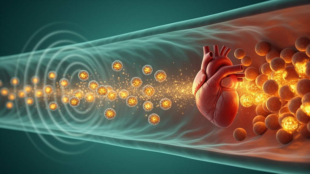
To solve the delivery dilemma, the team engineered a multifunctional nanoplatform called TEA@PLGA-PCM. The particles are built from poly(lactic-co-glycolic acid), or PLGA, a biocompatible and biodegradable copolymer used in FDA-approved products, loaded with TEA and a perfluorohexane core using a double-emulsion solvent evaporation method. The surface was decorated with PCM, a 20-amino-acid peptide with the sequence WLSEAGPVVTVRALRGTGSW that binds with high affinity to markers preferentially expressed on cardiomyocytes, especially under injury or stress. The final layer of control comes from low-intensity focused ultrasound, or LIFU, a non-invasive energy source that vaporizes the perfluorohexane core at the target site, triggering on-demand release of the drug only where the beam is pointed.
Characterization showed the chemistry worked as designed. Confocal microscopy confirmed successful PCM conjugation, and dynamic light scattering revealed that adding the peptide increased the average hydrodynamic diameter from 237.1 plus or minus 4.0 nanometers for TEA@PLGA to 286.8 plus or minus 7.0 nanometers for the targeted version, with the zeta potential shifting to approximately minus 34.7 plus or minus 3.3 millivolts. The targeted nanoparticles achieved a drug loading capacity of 5.7 plus or minus 1.3 percent and an encapsulation efficiency of 79.4 plus or minus 3.9 percent, and released roughly 40 percent of their TEA payload cumulatively over 48 hours in dialysis experiments. In H9c2 cells, flow cytometry showed that PCM decoration markedly increased nanoparticle uptake compared with non-targeted particles.
Before testing the platform, the researchers dissected the role of GPX4 in the injury process. In H9c2 cells exposed to ionizing radiation, irradiation significantly decreased cell viability, increased intracellular reactive oxygen species and raised levels of malondialdehyde, a lipid peroxidation marker. The classic ferroptosis inhibitor ferrostatin-1 rescued viability and reversed the ROS burst, while the ferroptosis inducer erastin worsened the damage, supporting ferroptosis as the operative cell death mechanism. When the team knocked down GPX4 with small interfering RNA, ROS accumulation, lipid peroxidation and loss of mitochondrial membrane potential all worsened, and protein analysis showed reduced Nrf2, SLC7A11, NQO1 and HO-1 alongside elevated NOX4 and KEAP1. Conversely, overexpressing GPX4 markedly suppressed the radiation-induced abnormalities. TEA intervention partially reversed the damage in both settings, consistent with the pathway hypothesis.
The in vivo experiments used C57BL/6 mice, eight to ten weeks old, given a single localized 20 Gy thoracic dose of X-ray radiation with the rest of the body shielded, then randomly assigned to eight treatment groups of ten animals each, ranging from oral free TEA at 150 milligrams per kilogram to intravenous nanoparticle formulations at 10 milligrams per kilogram, with or without LIFU exposure after each injection, plus a ferrostatin-1 comparator group. Over a four-week treatment period, biodistribution imaging showed that PCM-modified nanoparticles accumulated in the heart far better than unmodified ones, and that LIFU further enhanced cardiac enrichment, with the targeted, ultrasound-triggered group displaying the strongest fluorescence signal.
Functional outcomes followed the same pattern. Echocardiography revealed that irradiated mice suffered significantly impaired systolic function, with marked reductions in ejection fraction and fractional shortening, along with pathological remodeling including increased left ventricular mass and enlarged end-systolic and end-diastolic volumes. Free TEA partially restored function, the nanoparticle formulations performed better, and TEA@PLGA-PCM combined with LIFU showed the most pronounced protection, with ejection fraction and fractional shortening approaching control values and near-complete reversal of structural abnormalities, an effect comparable to the ferroptosis inhibitor group. Serum biomarkers reinforced the picture: radiation sharply elevated cardiac troponin I, a specific indicator of cardiomyocyte injury, and soluble ST2, which reflects myocardial strain and fibrosis, and the targeted ultrasound-triggered treatment significantly reduced both.
Molecular analysis of heart tissue linked the functional recovery to ferroptosis suppression through the proposed axis. Radiation elevated cardiac MDA levels and the NADP+/NADPH and GSSG/GSH ratios, all markers of oxidative stress and lipid peroxidation, while immunohistochemistry, immunofluorescence and western blotting showed downregulation of Nrf2, SLC7A11, GPX4, NQO1 and HO-1 and upregulation of NOX4 and KEAP1. TEA@PLGA-PCM with LIFU most effectively normalized these markers, matching the ferrostatin-1 positive control. The authors note an intriguing nuance: under severe oxidative stress the usual protective NRF2-NOX4 feedback loop appears disrupted, with radiation paradoxically lowering NRF2 while raising NOX4, possibly because excessive ROS impairs NRF2 function while NOX4 is activated through independent routes.
The researchers are careful about the boundaries of the work. The study focused on the cardiomyocyte-autonomous ferroptosis pathway and did not examine radiation-induced endothelial dysfunction, immune cell infiltration or fibroblast activation; the efficacy data are short-term, and long-term studies are needed to assess durability and nanoparticle biocompatibility after repeated dosing; and the team did not directly assess NRF2 nuclear translocation or its binding interaction with KEAP1. All results come from mouse models and H9c2 cells, so clinical translation remains unproven. Still, the authors argue the platform represents a promising strategy for integrated targeted delivery and synergistic therapy in RIHD management, potentially widening the therapeutic window of thoracic radiotherapy by protecting the heart without compromising tumor radiosensitivity.
Subject of Research: A targeted ultrasound-responsive nanoparticle delivering Abelmoschus manihot extract to inhibit radiation-induced cardiomyocyte ferroptosis via the NOX4/SLC7A11/GPX4 axis
Article Title: Ultrasound-targeted delivery of total extracts from Abelmoschus Manihot (L.) alleviates radiation-induced cardiomyocyte ferroptosis via the NOX4/SLC7A11/GPX4 axis
Article References: Niu, R., Xu, Z., Yang, W., Bao, T., Xu, F., Zhou, W., Zhang, L., Jia, Q., Wang, X., Chen, J., & Lin, X. (2026). Ultrasound-targeted delivery of total extracts from Abelmoschus Manihot (L.) alleviates radiation-induced cardiomyocyte ferroptosis via the NOX4/SLC7A11/GPX4 axis. Food Production, Processing and Nutrition, 8(1), Article 72. https://doi.org/10.1186/s43014-026-00422-8
Image Credits: AI Generated
DOI: 10.1186/s43014-026-00422-8
Keywords: radiation-induced heart disease, ferroptosis, nanoparticles, ultrasound-triggered drug delivery, Abelmoschus manihot, GPX4, NOX4, SLC7A11, cardioprotection, radiotherapy, PLGA, preclinical study
News Source: Nathaniel Bowman. (October 29, 2026). Ultrasound-Triggered Nanoparticles Deliver Herbal Extract to Shield Irradiated Hearts. Scienmag.



